Nigalye Archana K, Hess Kristina, Pundlik Shrinivas J, Jeffrey Brett G, Cukras Catherine A, Husain Deeba
Retina Service, Massachusetts Eye and Ear, Department of Ophthalmology, Harvard Medical School, 243 Charles St., Boston, MA 02114, USA.
National Eye Institute, National Institutes of Health, Bethesda, MD 20892, USA.
J Clin Med. 2022 Mar 1;11(5):1358. doi: 10.3390/jcm11051358.
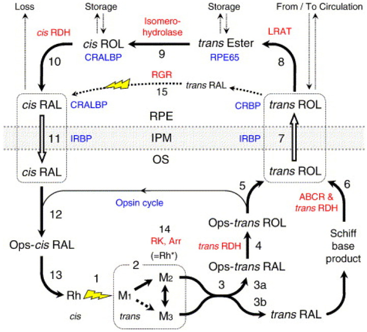
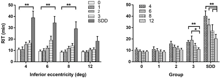
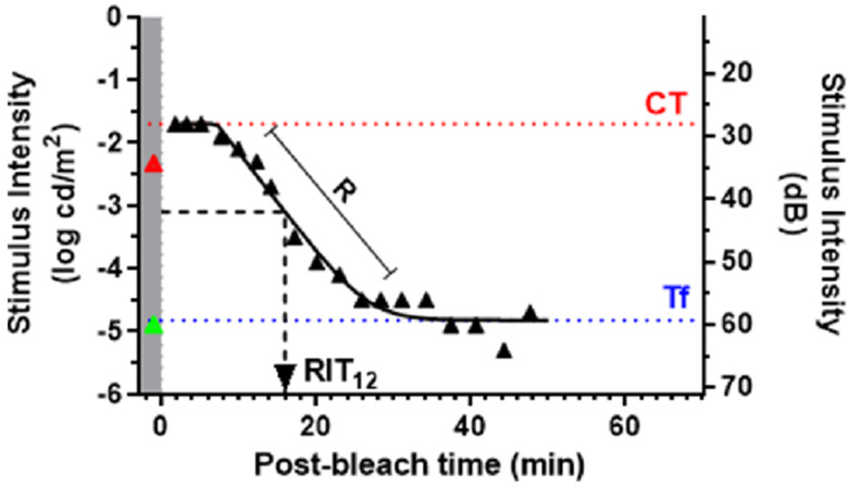
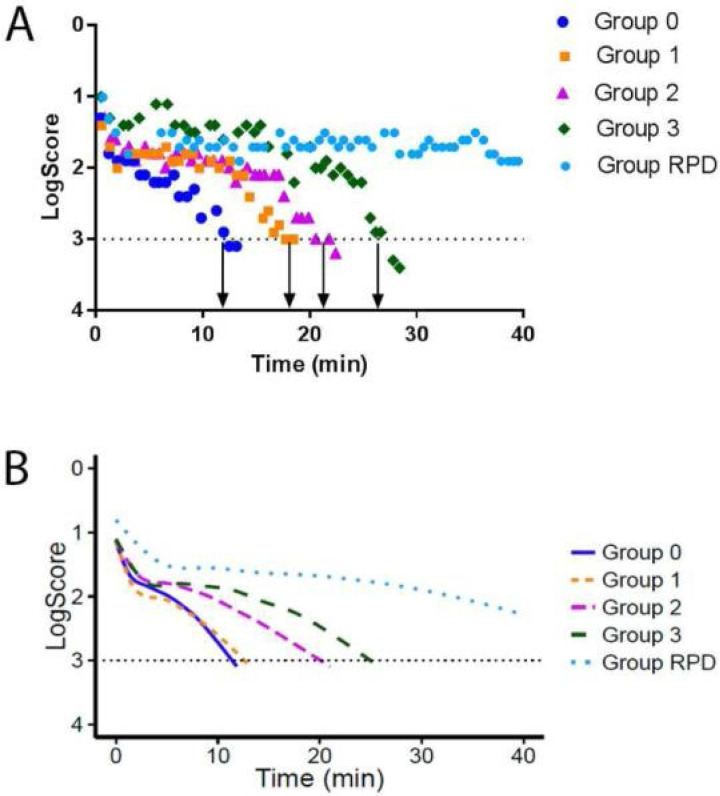
Dark adaptation (DA) refers to the slow recovery of visual sensitivity in darkness following exposure to intense or prolonged illumination, which bleaches a significant amount of the rhodopsin. This natural process also offers an opportunity to understand cellular function in the outer retina and evaluate for presence of disease. How our eyes adapt to darkness can be a key indicator of retinal health, which can be altered in the presence of certain diseases, such as age-related macular degeneration (AMD). A specific focus on clinical aspects of DA measurement and its significance to furthering our understanding of AMD has revealed essential findings underlying the pathobiology of the disease. The process of dark adaptation involves phototransduction taking place mainly between the photoreceptor outer segments and the retinal pigment epithelial (RPE) layer. DA occurs over a large range of luminance and is modulated by both cone and rod photoreceptors. In the photopic ranges, rods are saturated and cone cells adapt to the high luminance levels. However, under scotopic ranges, cones are unable to respond to the dim luminance and rods modulate the responses to lower levels of light as they can respond to even a single photon. Since the cone visual cycle is also based on the Muller cells, measuring the impairment in rod-based dark adaptation is thought to be particularly relevant to diseases such as AMD, which involves both photoreceptors and RPE. Dark adaptation parameters are metrics derived from curve-fitting dark adaptation sensitivities over time and can represent specific cellular function. Parameters such as the cone-rod break (CRB) and rod intercept time (RIT) are particularly sensitive to changes in the outer retina. There is some structural and functional continuum between normal aging and the AMD pathology. Many studies have shown an increase of the rod intercept time (RIT), i.e., delays in rod-mediated DA in AMD patients with increasing disease severity determined by increased drusen grade, pigment changes and the presence of subretinal drusenoid deposits (SDD) and association with certain morphological features in the peripheral retina. Specifications of spatial testing location, repeatability of the testing, ease and availability of the testing device in clinical settings, and test duration in elderly population are also important. We provide a detailed overview in light of all these factors.
暗适应(DA)是指在暴露于强光或长时间光照后,在黑暗中视觉敏感度的缓慢恢复,这种光照会使大量视紫红质褪色。这一自然过程也为了解视网膜外层的细胞功能和评估疾病的存在提供了机会。我们的眼睛如何适应黑暗可能是视网膜健康的关键指标,在某些疾病(如年龄相关性黄斑变性(AMD))存在时,这一指标可能会发生改变。对暗适应测量的临床方面及其对加深我们对AMD理解的意义的特别关注,揭示了该疾病病理生物学的重要发现。暗适应过程涉及主要发生在光感受器外段和视网膜色素上皮(RPE)层之间的光转导。暗适应发生在大范围的亮度范围内,并受到视锥和视杆光感受器的调节。在明视觉范围内,视杆细胞饱和,视锥细胞适应高亮度水平。然而,在暗视觉范围内,视锥细胞无法对昏暗的亮度做出反应,视杆细胞调节对较低光水平的反应,因为它们甚至可以对单个光子做出反应。由于视锥细胞视觉循环也基于米勒细胞,因此测量基于视杆细胞的暗适应损伤被认为与AMD等疾病特别相关,AMD涉及光感受器和RPE。暗适应参数是通过对暗适应敏感度随时间进行曲线拟合得出的指标,可以代表特定的细胞功能。诸如视锥-视杆转折点(CRB)和视杆截距时间(RIT)等参数对外层视网膜的变化特别敏感。正常衰老和AMD病理之间存在一些结构和功能上的连续性。许多研究表明,视杆截距时间(RIT)增加,即AMD患者中视杆介导的暗适应延迟,疾病严重程度增加,这由玻璃膜疣等级增加、色素变化以及视网膜下玻璃膜疣样沉积物(SDD)的存在以及与周边视网膜某些形态特征的关联来确定。空间测试位置的规格、测试的可重复性、临床环境中测试设备的易用性和可用性以及老年人群的测试持续时间也很重要。我们根据所有这些因素提供详细概述。