Department of Radiation Oncology, Fudan University Shanghai Cancer Center, 270 DongAn Road, Shanghai, 200032, China.
Department of Radiology, Fudan University Shanghai Cancer Center, Shanghai, 200032, China.
Breast Cancer Res. 2022 Mar 15;24(1):20. doi: 10.1186/s13058-022-01516-0.
This study investigated the efficacy of radiomics to predict survival outcome for locally advanced breast cancer (LABC) patients and the association of radiomics with tumor heterogeneity and microenvironment.
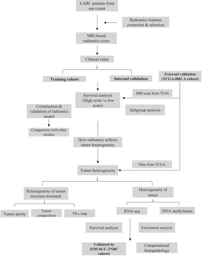
Patients with LABC from 2010 to 2015 were retrospectively reviewed. Radiomics features were extracted from enhanced MRI. We constructed the radiomics score using lasso and assessed its prognostic value. An external validation cohort from The Cancer Imaging Archive was used to assess phenotype reproducibility. Sequencing data from TCGA and our center were applied to reveal genomic landscape of different radiomics score groups. Tumor infiltrating lymphocytes map and bioinformatics methods were applied to evaluate the heterogeneity of tumor microenvironment. Computational histopathology was also applied.
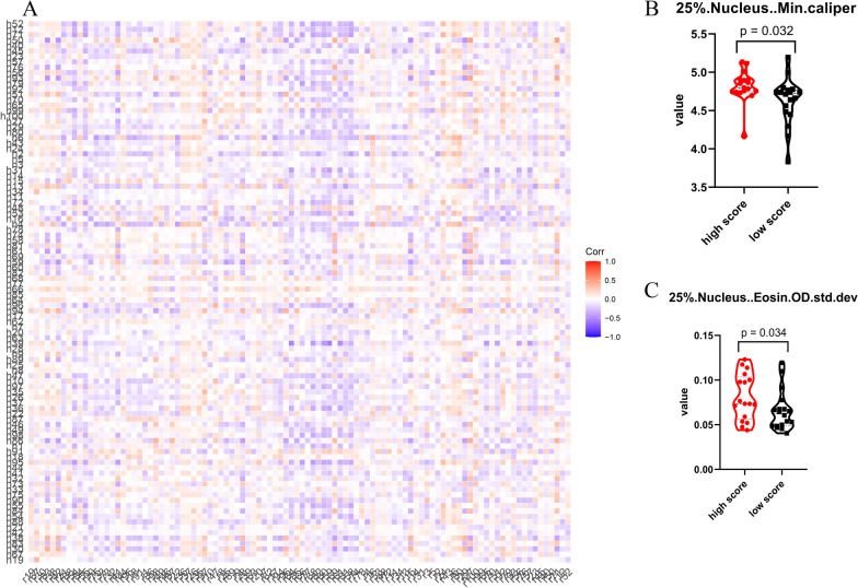
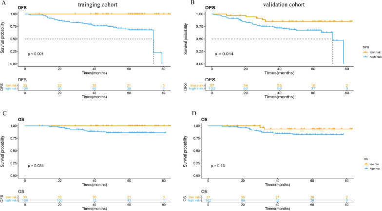
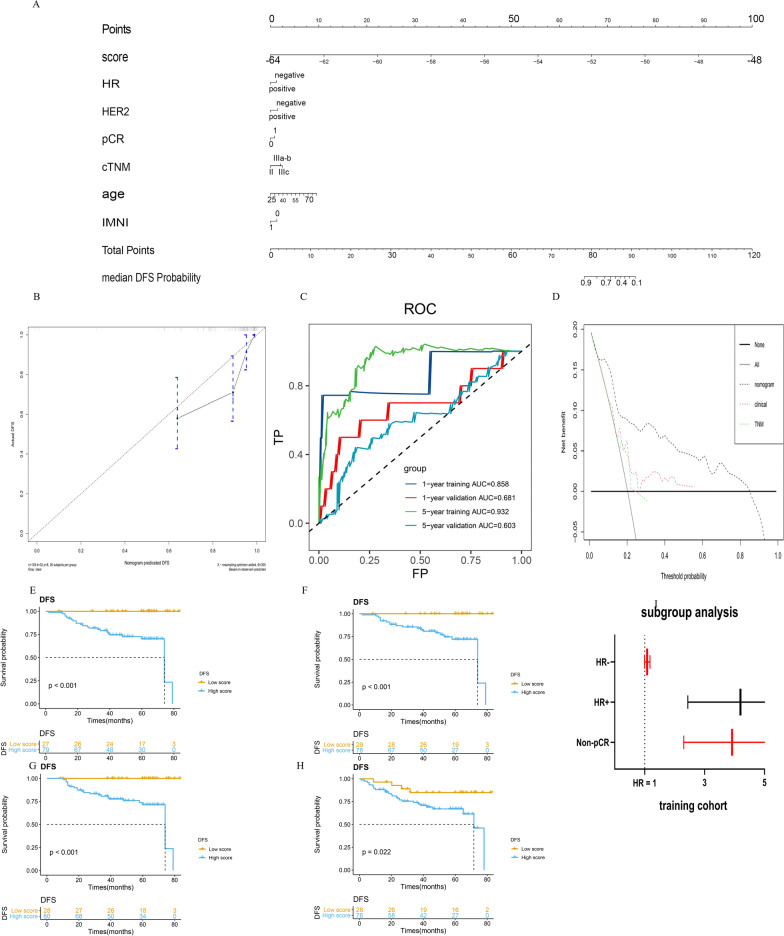
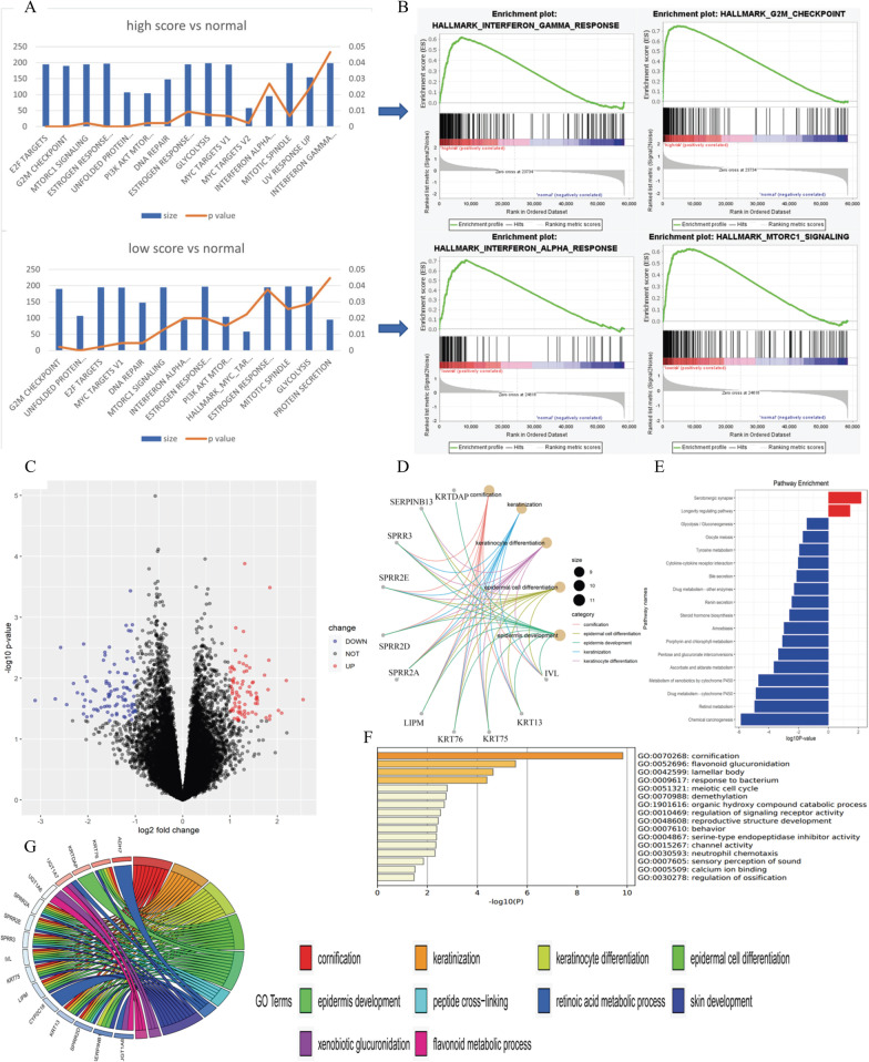
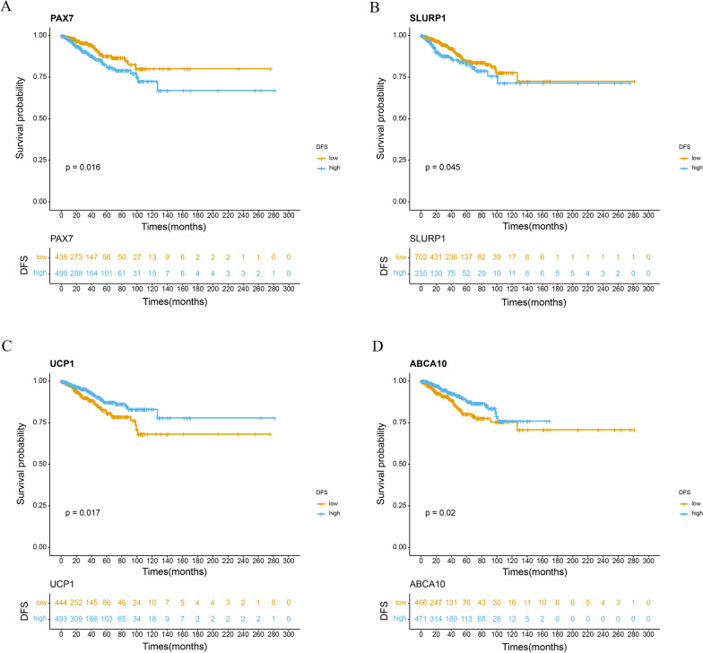
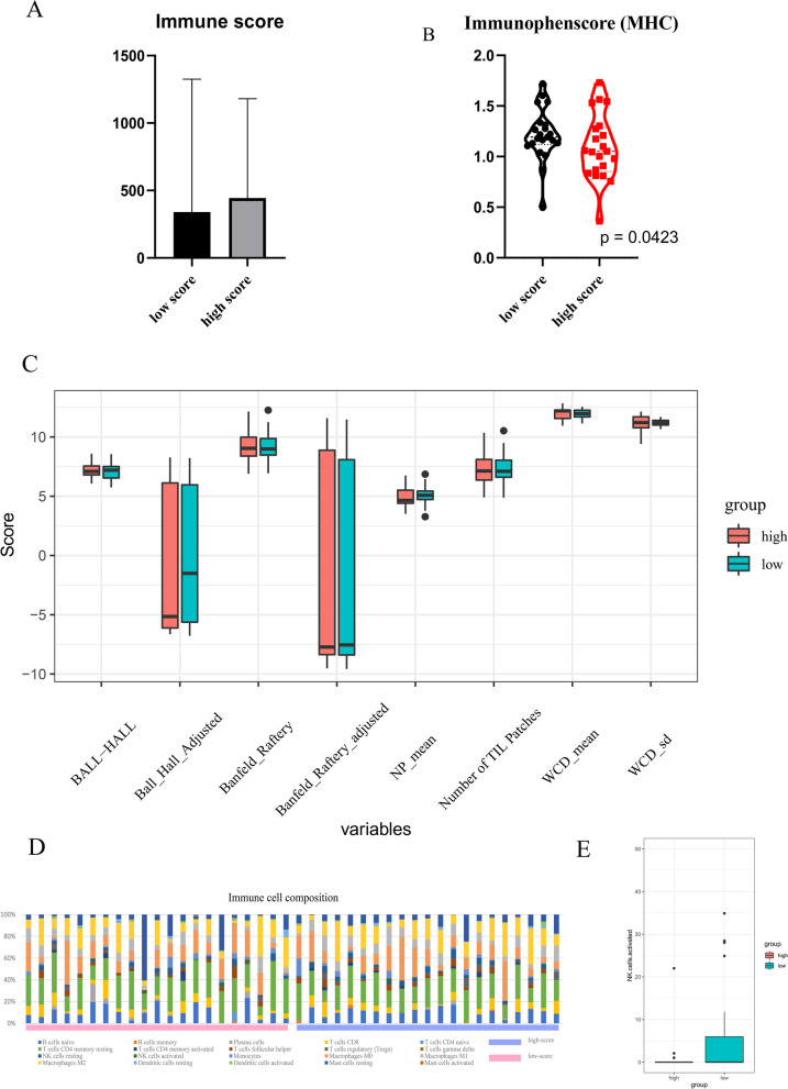
A total of 278 patients were divided into training cohort and validation cohort. Radiomics score was constructed and significantly associated with disease-free survival (DFS) of the patients in training cohort, validation cohort and external validation cohort (p < 0.001, p = 0.014 and p = 0.041, respectively). The radiomics-based nomogram showed better predictive performance of DFS compared with TNM model. Distinct gene expression patterns were identified. Immunophenotype and immune cell composition was different in each radiomics score group. The link between radiomics and computational histopathology was revealed.
The radiomics score could effectively predict prognosis of LABC after neoadjuvant chemotherapy and radiotherapy. Radiomics revealed heterogeneity of tumor cell and tumor microenvironment and holds great potential to facilitate individualized DFS estimation and guide personalized care.
本研究旨在探讨影像组学预测局部晚期乳腺癌(LABC)患者生存结局的疗效,以及影像组学与肿瘤异质性和微环境的关系。
回顾性分析 2010 年至 2015 年期间的 LABC 患者。从增强 MRI 中提取影像组学特征。使用lasso 构建影像组学评分,并评估其预后价值。使用癌症影像学档案的外部验证队列评估表型重现性。应用 TCGA 和我们中心的测序数据揭示不同影像组学评分组的基因组图谱。肿瘤浸润淋巴细胞图谱和生物信息学方法用于评估肿瘤微环境的异质性。还应用了计算组织病理学。
共 278 例患者分为训练队列和验证队列。构建了影像组学评分,并与训练队列、验证队列和外部验证队列患者的无病生存(DFS)显著相关(p<0.001,p=0.014 和 p=0.041)。基于影像组学的列线图显示出比 TNM 模型更好的 DFS 预测性能。确定了不同的基因表达模式。在每个影像组学评分组中,免疫表型和免疫细胞组成不同。揭示了影像组学与计算组织病理学之间的联系。
影像组学评分可有效预测新辅助化疗和放疗后 LABC 的预后。影像组学揭示了肿瘤细胞和肿瘤微环境的异质性,具有促进个体化 DFS 估计和指导个体化护理的巨大潜力。