Department of Ophthalmology, Bascom Palmer Eye Institute, University of Miami Miller, School of Medicine, Miami, USA; Department of Ophthalmology and Visual Science, Yale School of Medicine, New Haven, CT, USA.
Department of Ophthalmology, Bascom Palmer Eye Institute, University of Miami Miller, School of Medicine, Miami, USA; Ophthalmic Center, The Second Affiliated Hospital of Guangzhou Medical University, Guangzhou, Guangdong, China.
Ocul Surf. 2022 Jul;25:8-18. doi: 10.1016/j.jtos.2022.03.006. Epub 2022 Mar 29.
Optical coherence tomography angiography (OCTA) was utilized to examine changes in ocular surface squamous neoplasia (OSSN) vascular patterns over time in individuals treated with topical medical therapy.
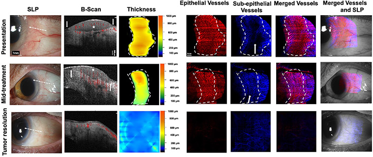
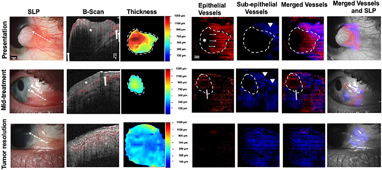
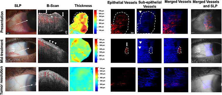
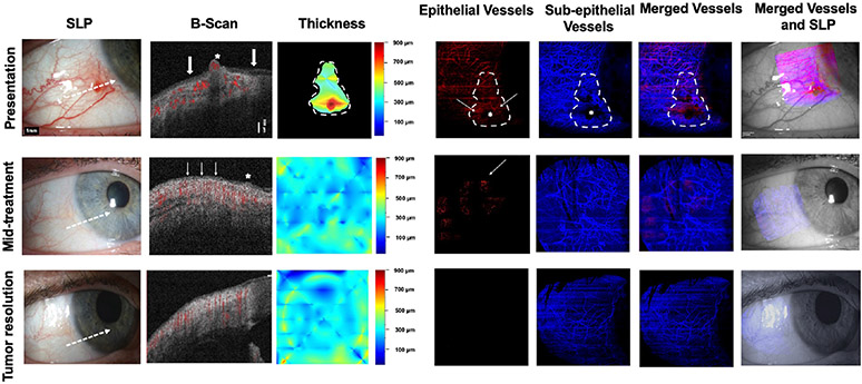
Ten individuals with OSSN diagnosed by clinical examination and high resolution (HR)-optical coherence tomography (OCT) were recruited. All individuals received topical immuno- or chemotherapy. OCTA images were obtained and analyzed at three points: presentation, mid-treatment and tumor resolution. Tumor metrics including tumor area (TA), tumor volume (TV), tumor depth (TD), and total tumor density (TTD) were calculated. Vessel area density (VAD) was also quantified within the OSSN, the subepithelium under and adjacent to the OSSN and the subepithelium of the uninvolved, contralateral eye. Vascular network changes were also subjectively evaluated.
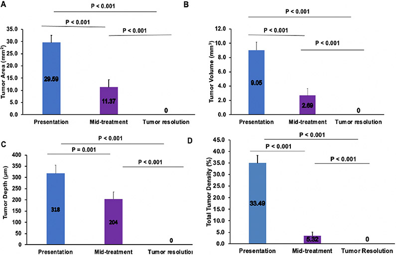
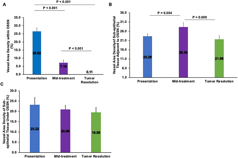
TA, TV, TD and TTD all significantly decreased with time (p < 0.001). The mean VAD within the OSSN significantly decreased (p < 0.001) between visits (presentation: 26.52 ± 6.8%, mid-treatment: 7.19 ± 5.88%, tumor resolution: 0.11 ± 0.34%). The mean subepithelial VAD under the OSSN also decreased with time (23.22 ± 11.03%, 20.99 ± 5.99% and 19.58 ± 7.08%), and after resolution the sub-tumor VAD (19.58 ± 7.08%) was comparable to the subepithelial VAD in the contralateral eye (15.47 ± 4.37%, p > 0.05). The mean VAD in the subepithelium adjacent to the OSSN increased with treatment, then decreased significantly between mid-treatment and resolution (23.26 ± 4.54, 28.30 ± 7.43% and 21.68 ± 6.10%, p = 0.009). Qualitatively, the tumor subepithelial vascular network was complex and dense but with tumor resolution appeared less tortuous and similar to the uninvolved eye.
OCTA provided insight into the pathophysiology of tumor angiogenesis, showing decreased vascular density and normalization of vascular networks associated with tumor resolution.
利用光相干断层扫描血管造影术(OCTA)观察经局部药物治疗的眼表鳞状细胞癌(OSSN)患者的眼表病变血管模式随时间的变化。
招募了 10 名经临床检查和高分辨率(HR)光学相干断层扫描(OCT)诊断为 OSSN 的患者。所有患者均接受局部免疫或化学治疗。在三个时间点:就诊时、治疗中期和肿瘤消退时,获得 OCTA 图像并进行分析。计算肿瘤指标,包括肿瘤面积(TA)、肿瘤体积(TV)、肿瘤深度(TD)和总肿瘤密度(TTD)。还定量评估了 OSSN 下、OSSN 下和相邻上皮下的血管面积密度(VAD)以及对侧未受累眼的上皮下血管密度。还对血管网络变化进行了主观评估。
TA、TV、TD 和 TTD 均随时间显著降低(p<0.001)。OSSN 内的平均 VAD 在就诊时(26.52±6.8%)、治疗中期(7.19±5.88%)和肿瘤消退时(0.11±0.34%)显著降低(p<0.001)。OSSN 下的平均上皮下 VAD 也随时间减少(23.22±11.03%、20.99±5.99%和 19.58±7.08%),消退后肿瘤下 VAD(19.58±7.08%)与对侧眼的上皮下 VAD(15.47±4.37%)相当(p>0.05)。OSSN 相邻上皮下的平均 VAD 在治疗过程中增加,然后在治疗中期和消退时显著降低(23.26±4.54、28.30±7.43%和 21.68±6.10%,p=0.009)。定性地说,肿瘤上皮下血管网络复杂而密集,但随着肿瘤消退,血管网络变得不那么扭曲,与未受累眼相似。
OCTA 提供了肿瘤血管生成病理生理学的深入了解,显示血管密度降低,与肿瘤消退相关的血管网络正常化。