Kos Mark Z, Puppala Sobha, Cruz Dianne, Neary Jennifer L, Kumar Ashish, Dalan Emma, Li Cun, Nathanielsz Peter, Carless Melanie A
Department of Human Genetics, South Texas Diabetes and Obesity Institute, University of Texas Rio Grande Valley School of Medicine, Edinburg, TX, United States.
Department of Internal Medicine-Section of Molecular Medicine, Wake Forest Baptist Medical Center, Winston-Salem, NC, United States.
Front Mol Neurosci. 2022 Mar 22;15:817290. doi: 10.3389/fnmol.2022.817290. eCollection 2022.
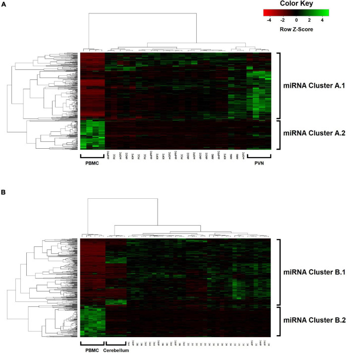
The use of easily accessible peripheral samples, such as blood or saliva, to investigate neurological and neuropsychiatric disorders is well-established in genetic and epigenetic research, but the pathological implications of such biomarkers are not easily discerned. To better understand the relationship between peripheral blood- and brain-based epigenetic activity, we conducted a pilot study on captive baboons () to investigate correlations between miRNA expression in peripheral blood mononuclear cells (PBMCs) and 14 different cortical and subcortical brain regions, represented by two study groups comprised of 4 and 6 animals. Using next-generation sequencing, we identified 362 miRNAs expressed at ≥ 10 read counts in 80% or more of the brain samples analyzed. Nominally significant pairwise correlations (one-sided < 0.05) between peripheral blood and mean brain expression levels of individual miRNAs were observed for 39 and 44 miRNAs in each group. When miRNA expression levels were averaged for tissue type across animals within the groups, Spearman's rank correlations between PBMCs and the brain regions are all highly significant ( = 0.47-0.57; < 2.2 × 10), although pairwise correlations among the brain regions are markedly stronger ( = 0.86-0.99). Principal component analysis revealed differentiation in miRNA expression between peripheral blood and the brain regions for the first component (accounting for ∼75% of variance). Linear mixed effects modeling attributed most of the variance in expression to differences between miRNAs (>70%), with non-significant 7.5% and 13.1% assigned to differences between blood and brain-based samples in the two study groups. Hierarchical UPGMA clustering revealed a major co-expression branch in both study groups, comprised of miRNAs globally upregulated in blood relative to the brain samples, exhibiting an enrichment of miRNAs expressed in immune cells (CD14+, CD15+, CD19+, CD3+, and CD56 + leukocytes) among the top blood-brain correlates, with the gene , encoding a master transcription factor that regulates angiogenesis and neural stem cell activation, representing the most prevalent miRNA target. Although some differentiation was observed between tissue types, these preliminary findings reveal wider correlated patterns between blood- and brain-expressed miRNAs, suggesting the potential utility of blood-based miRNA profiling for investigating by proxy certain miRNA activity in the brain, with implications for neuroinflammatory and c-Myc-mediated processes.
在遗传和表观遗传学研究中,利用易于获取的外周样本(如血液或唾液)来研究神经和神经精神疾病已得到广泛应用,但此类生物标志物的病理意义并不容易识别。为了更好地理解外周血与脑源性表观遗传活性之间的关系,我们对圈养狒狒开展了一项初步研究,以调查外周血单核细胞(PBMC)中miRNA表达与14个不同皮质和皮质下脑区之间的相关性,研究分为两个组,分别包含4只和6只动物。通过新一代测序,我们在80%或更多分析的脑样本中鉴定出表达量≥10 reads的362种miRNA。每组中分别有39种和44种miRNA在外周血与单个miRNA的平均脑表达水平之间观察到名义上显著的成对相关性(单侧P<0.05)。当对组内动物的组织类型平均miRNA表达水平时,PBMC与脑区之间的Spearman等级相关性均高度显著(r = 0.47 - 0.57;P < 2.2×10),尽管脑区之间的成对相关性明显更强(r = 0.86 - 0.99)。主成分分析显示第一成分在外周血和脑区的miRNA表达上存在差异(占方差的约75%)。线性混合效应模型将大部分表达差异归因于miRNA之间的差异(>70%),两个研究组中分别有7.5%和13.1%的差异归因于血源性样本和脑源性样本之间的差异,差异不显著。层次UPGMA聚类显示两个研究组中均有一个主要的共表达分支,由相对于脑样本在血液中整体上调的miRNA组成,在血脑相关性最强的miRNA中,免疫细胞(CD14 +、CD15 +、CD19 +、CD3 +和CD56 +白细胞)中表达的miRNA富集,编码调节血管生成和神经干细胞激活的主转录因子的基因是最常见的miRNA靶标。尽管在组织类型之间观察到了一些差异,但这些初步发现揭示了血源性和脑源性miRNA之间更广泛的相关模式,表明基于血液的miRNA谱分析可能有助于间接研究大脑中的某些miRNA活性,对神经炎症和c-Myc介导的过程具有重要意义。