• 文献检索
  • 文档翻译
  • 深度研究
  • 学术资讯
  • Suppr Zotero 插件Zotero 插件
  • 邀请有礼
  • 套餐&价格
  • 历史记录
应用&插件
Suppr Zotero 插件Zotero 插件浏览器插件Mac 客户端Windows 客户端微信小程序
定价
高级版会员购买积分包购买API积分包
服务
文献检索文档翻译深度研究API 文档MCP 服务
关于我们
关于 Suppr公司介绍联系我们用户协议隐私条款
关注我们

Suppr 超能文献

核心技术专利:CN118964589B侵权必究
粤ICP备2023148730 号-1Suppr @ 2026

文献检索

告别复杂PubMed语法,用中文像聊天一样搜索,搜遍4000万医学文献。AI智能推荐,让科研检索更轻松。

立即免费搜索

文件翻译

保留排版,准确专业,支持PDF/Word/PPT等文件格式,支持 12+语言互译。

免费翻译文档

深度研究

AI帮你快速写综述,25分钟生成高质量综述,智能提取关键信息,辅助科研写作。

立即免费体验

基于分水岭预处理的区域生长法对CT上自发性脑出血的分割

Segmentation of Spontaneous Intracerebral Hemorrhage on CT With a Region Growing Method Based on Watershed Preprocessing.

作者信息

Zhou Zhengsong, Wan Hongli, Zhang Haoyu, Chen Xumiao, Wang Xiaoyu, Lili Shiluo, Zhang Tao

机构信息

Department of Electronic Information Engineering, Chengdu Jincheng College, Chengdu, China.

Department of Epidemiology and Health Statistics, West China School of Public Health and West China Fourth Hospital, Sichuan University, Chengdu, China.

出版信息

Front Neurol. 2022 Mar 29;13:865023. doi: 10.3389/fneur.2022.865023. eCollection 2022.

DOI:10.3389/fneur.2022.865023
PMID:35422751
原文链接:https://pmc.ncbi.nlm.nih.gov/articles/PMC9002175/
Abstract

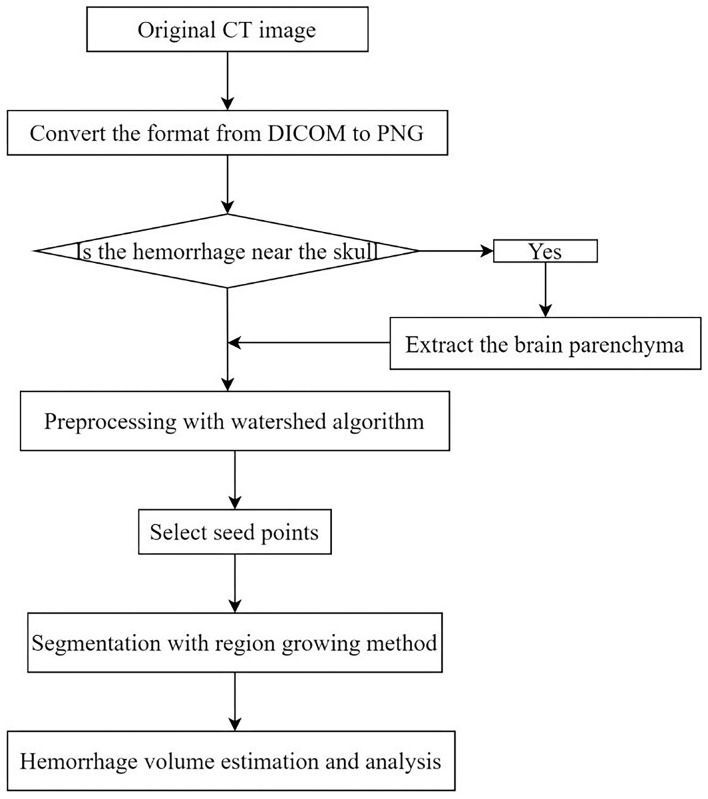
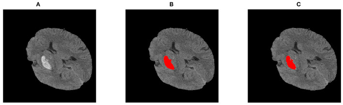
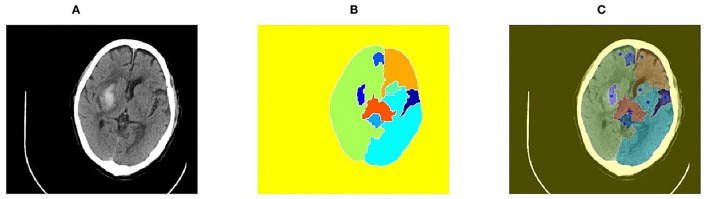
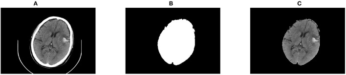
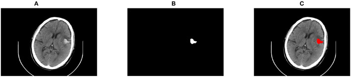
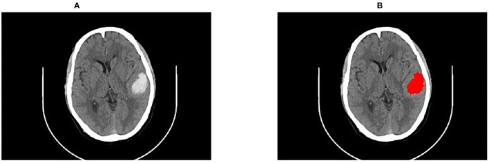
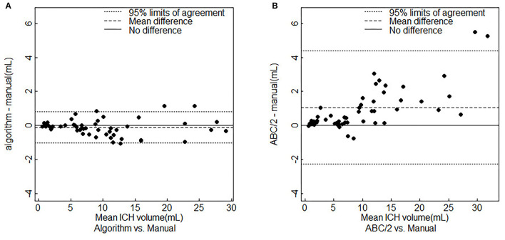
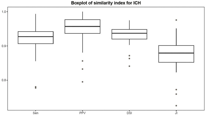
Intracerebral hemorrhage (ICH) poses a great threat to human life due to its high incidence and poor prognosis. Identification of the bleeding location and quantification of the volume based on CT images are of great significance for assisting the diagnosis and treatment of ICH. In this study, a region-growing algorithm based on watershed preprocessing (RG-WP) was proposed to segment and quantify the hemorrhage. The lowest points yielded by the watershed algorithm were used as seed points for region growing and then hemorrhage was segmented based on the region growing method. At the same time, to integrate the rich experience of clinicians with the algorithm, manual selection of seed points on the basis of watershed segmentation was performed. With the application of segmentation on CT images of 55 patients with ICH, the performance of the RG-WP algorithm was evaluated by comparing it with manual segmentations delineated by professional clinicians as well as the traditional ABC/2 method and the deep learning algorithm U-net. The mean deviation of hemorrhage volume of the RG-WP algorithm from manual segmentation was -0.12 ml (range: -1.05-1.16), while that of the ABC/2 from the manual was 1.05 ml (range: -0.77-9.57). Strong agreement of the algorithm and the manual was confirmed with a high intraclass correlation coefficient (ICC) (0.998, 95% : 0.997-0.999), which was superior to that of the ABC/2 and the manual (0.972, 95% : 0.953-0.984). The sensitivity (Sen), positive predictive value (PPV), dice similarity index (DSI), and Jaccard index (JI) of the RG-WP algorithm compared to the manual were 0.92 ± 0.04, 0.95 ± 0.04, 0.93 ± 0.02, and 0.88 ± 0.04, respectively, showing high consistency. Besides, the accuracy of the algorithm was also comparable to that of the deep learning method U-net, with Sen, PPV, DSI, and JI being 0.91 ± 0.09, 0.91 ± 0.06, 0.91 ± 0.05, and 0.91 ± 0.06, respectively.

摘要

脑出血(ICH)因其高发病率和不良预后对人类生命构成巨大威胁。基于CT图像识别出血位置并对出血量进行量化,对于辅助脑出血的诊断和治疗具有重要意义。在本研究中,提出了一种基于分水岭预处理的区域生长算法(RG-WP)来分割和量化出血区域。将分水岭算法产生的最低点用作区域生长的种子点,然后基于区域生长方法分割出血区域。同时,为了将临床医生的丰富经验与该算法相结合,在分水岭分割的基础上进行了种子点的手动选择。通过对55例脑出血患者的CT图像进行分割应用,将RG-WP算法与专业临床医生手动勾勒的分割结果、传统的ABC/2方法以及深度学习算法U-net进行比较,评估了RG-WP算法的性能。RG-WP算法与手动分割的出血量平均偏差为-0.12 ml(范围:-1.05至1.16),而ABC/2与手动分割的平均偏差为1.05 ml(范围:-0.77至9.57)。通过高组内相关系数(ICC)(0.998,95%:0.997 - 0.999)证实了该算法与手动分割有很强的一致性,优于ABC/2与手动分割的一致性(0.972,95%:0.953 - 0.984)。与手动分割相比,RG-WP算法的灵敏度(Sen)、阳性预测值(PPV)、骰子相似性指数(DSI)和杰卡德指数(JI)分别为0.92±0.04、0.95±0.04、0.93±0.02和0.88±0.04,显示出高度一致性。此外,该算法的准确性也与深度学习方法U-net相当,Sen、PPV、DSI和JI分别为0.91±0.09、0.91±0.06、0.91±0.05和0.91±0.06。

https://cdn.ncbi.nlm.nih.gov/pmc/blobs/dc60/9002175/c6e0dcf65a55/fneur-13-865023-g0008.jpg
https://cdn.ncbi.nlm.nih.gov/pmc/blobs/dc60/9002175/54a49138502e/fneur-13-865023-g0001.jpg
https://cdn.ncbi.nlm.nih.gov/pmc/blobs/dc60/9002175/1b08a241ca1d/fneur-13-865023-g0002.jpg
https://cdn.ncbi.nlm.nih.gov/pmc/blobs/dc60/9002175/22a662b5f7d3/fneur-13-865023-g0003.jpg
https://cdn.ncbi.nlm.nih.gov/pmc/blobs/dc60/9002175/c8bad9c53491/fneur-13-865023-g0004.jpg
https://cdn.ncbi.nlm.nih.gov/pmc/blobs/dc60/9002175/81e536078283/fneur-13-865023-g0005.jpg
https://cdn.ncbi.nlm.nih.gov/pmc/blobs/dc60/9002175/5d9c3e3475ea/fneur-13-865023-g0006.jpg
https://cdn.ncbi.nlm.nih.gov/pmc/blobs/dc60/9002175/9cb3838f92f6/fneur-13-865023-g0007.jpg
https://cdn.ncbi.nlm.nih.gov/pmc/blobs/dc60/9002175/c6e0dcf65a55/fneur-13-865023-g0008.jpg

相似文献

1
Segmentation of Spontaneous Intracerebral Hemorrhage on CT With a Region Growing Method Based on Watershed Preprocessing.基于分水岭预处理的区域生长法对CT上自发性脑出血的分割
Front Neurol. 2022 Mar 29;13:865023. doi: 10.3389/fneur.2022.865023. eCollection 2022.
2
[Hematoma Segmentation of Spontaneous Intracerebral Hemorrhage Based on Watershed and Region-Growing Algorithm].基于分水岭和区域生长算法的自发性脑出血血肿分割
Sichuan Da Xue Xue Bao Yi Xue Ban. 2022 May;53(3):511-516. doi: 10.12182/20220560202.
3
Efficiency of a deep learning-based artificial intelligence diagnostic system in spontaneous intracerebral hemorrhage volume measurement.基于深度学习的人工智能诊断系统在自发性脑出血容量测量中的效率。
BMC Med Imaging. 2021 Aug 13;21(1):125. doi: 10.1186/s12880-021-00657-6.
4
PItcHPERFeCT: Primary Intracranial Hemorrhage Probability Estimation using Random Forests on CT.PItcHPERFeCT:使用CT上的随机森林进行原发性颅内出血概率估计
Neuroimage Clin. 2017 Feb 15;14:379-390. doi: 10.1016/j.nicl.2017.02.007. eCollection 2017.
5
Development and Validation of an Automatic Segmentation Algorithm for Quantification of Intracerebral Hemorrhage.用于脑内出血定量的自动分割算法的开发与验证
Stroke. 2016 Nov;47(11):2776-2782. doi: 10.1161/STROKEAHA.116.013779. Epub 2016 Oct 4.
6
Deep Network for the Automatic Segmentation and Quantification of Intracranial Hemorrhage on CT.用于CT图像上颅内出血自动分割与量化的深度网络
Front Neurosci. 2021 Jan 11;14:541817. doi: 10.3389/fnins.2020.541817. eCollection 2020.
7
Semantic Segmentation of Spontaneous Intracerebral Hemorrhage, Intraventricular Hemorrhage, and Associated Edema on CT Images Using Deep Learning.利用深度学习对CT图像上的自发性脑出血、脑室内出血及相关水肿进行语义分割
Radiol Artif Intell. 2022 Sep 28;4(6):e220096. doi: 10.1148/ryai.220096. eCollection 2022 Nov.
8
Fully Automated Segmentation Algorithm for Hematoma Volumetric Analysis in Spontaneous Intracerebral Hemorrhage.全自动血肿体积分析算法在自发性脑出血中的应用。
Stroke. 2019 Dec;50(12):3416-3423. doi: 10.1161/STROKEAHA.119.026561. Epub 2019 Nov 18.
9
Deep learning-based computed tomography image segmentation and volume measurement of intracerebral hemorrhage.基于深度学习的脑出血计算机断层扫描图像分割与体积测量
Front Neurosci. 2022 Oct 3;16:965680. doi: 10.3389/fnins.2022.965680. eCollection 2022.
10
A symmetric prior knowledge based deep learning model for intracerebral hemorrhage lesion segmentation.一种基于对称先验知识的脑出血病灶分割深度学习模型。
Front Physiol. 2022 Nov 23;13:977427. doi: 10.3389/fphys.2022.977427. eCollection 2022.

引用本文的文献

1
Adapting foundation models for rapid clinical response: intracerebral hemorrhage segmentation in emergency settings.使基础模型适应快速临床响应:急诊环境下的脑出血分割
Sci Rep. 2025 Aug 3;15(1):28314. doi: 10.1038/s41598-025-13742-5.
2
[Research on intelligent tooth segmentation method combining multiple seed region growth and boundary extension].基于多种子区域生长与边界扩展相结合的智能牙齿分割方法研究
Sheng Wu Yi Xue Gong Cheng Xue Za Zhi. 2024 Jun 25;41(3):520-526. doi: 10.7507/1001-5515.202309030.
3
Accuracy of automated intracerebral hemorrhage volume measurement on non-contrast computed tomography: a Swedish Stroke Register cohort study.

本文引用的文献

1
U-Net: A valuable encoder-decoder architecture for liver tumors segmentation in CT images.U-Net:一种用于CT图像中肝脏肿瘤分割的有价值的编码器-解码器架构。
J Xray Sci Technol. 2022;30(1):45-56. doi: 10.3233/XST-210993.
2
Dual-Path Attention Compensation U-Net for Stroke Lesion Segmentation.双路径注意力补偿 U-Net 用于脑卒中病灶分割。
Comput Intell Neurosci. 2021 Aug 31;2021:7552185. doi: 10.1155/2021/7552185. eCollection 2021.
3
Management of Intracerebral Hemorrhage: JACC Focus Seminar.颅内出血的管理:美国心脏病学会焦点研讨会。
非对比 CT 自动测量脑出血量的准确性:一项瑞典卒中登记队列研究。
Neuroradiology. 2023 Mar;65(3):479-488. doi: 10.1007/s00234-022-03075-9. Epub 2022 Nov 3.
J Am Coll Cardiol. 2020 Apr 21;75(15):1819-1831. doi: 10.1016/j.jacc.2019.10.066.
4
DENSE-INception U-net for medical image segmentation.基于密集卷积 Inception 的 U-Net 网络在医学图像分割中的应用
Comput Methods Programs Biomed. 2020 Aug;192:105395. doi: 10.1016/j.cmpb.2020.105395. Epub 2020 Feb 15.
5
Fully Automated Segmentation Algorithm for Hematoma Volumetric Analysis in Spontaneous Intracerebral Hemorrhage.全自动血肿体积分析算法在自发性脑出血中的应用。
Stroke. 2019 Dec;50(12):3416-3423. doi: 10.1161/STROKEAHA.119.026561. Epub 2019 Nov 18.
6
Fast and fully-automated detection and segmentation of pulmonary nodules in thoracic CT scans using deep convolutional neural networks.使用深度卷积神经网络快速、全自动检测和分割胸部 CT 扫描中的肺结节。
Comput Med Imaging Graph. 2019 Jun;74:25-36. doi: 10.1016/j.compmedimag.2019.02.003. Epub 2019 Mar 22.
7
Improving Sensitivity on Identification and Delineation of Intracranial Hemorrhage Lesion Using Cascaded Deep Learning Models.使用级联深度学习模型提高颅内出血病变识别与描绘的敏感性
J Digit Imaging. 2019 Jun;32(3):450-461. doi: 10.1007/s10278-018-00172-1.
8
Intracerebral Hemorrhage.脑出血
Continuum (Minneap Minn). 2018 Dec;24(6):1603-1622. doi: 10.1212/CON.0000000000000672.
9
Computer-assisted delineation of hematoma from CT volume using autoencoder and Chan Vese model.基于自动编码器和 Chan-Vese 模型的 CT 容积图像血肿自动勾画。
Int J Comput Assist Radiol Surg. 2019 Feb;14(2):259-269. doi: 10.1007/s11548-018-1873-9. Epub 2018 Oct 30.
10
Convolutional neural networks: an overview and application in radiology.卷积神经网络:概述及其在放射学中的应用。
Insights Imaging. 2018 Aug;9(4):611-629. doi: 10.1007/s13244-018-0639-9. Epub 2018 Jun 22.