Muschelli John, Sweeney Elizabeth M, Ullman Natalie L, Vespa Paul, Hanley Daniel F, Crainiceanu Ciprian M
Department of Biostatistics, Bloomberg School of Public Health, Johns Hopkins University, Baltimore, MD, USA.
Department of Neurology, Division of Brain Injury Outcomes, Johns Hopkins Medical Institutions, Baltimore, MD, USA.
Neuroimage Clin. 2017 Feb 15;14:379-390. doi: 10.1016/j.nicl.2017.02.007. eCollection 2017.
Intracerebral hemorrhage (ICH), where a blood vessel ruptures into areas of the brain, accounts for approximately 10-15% of all strokes. X-ray computed tomography (CT) scanning is largely used to assess the location and volume of these hemorrhages. Manual segmentation of the CT scan using planimetry by an expert reader is the gold standard for volume estimation, but is time-consuming and has within- and across-reader variability. We propose a fully automated segmentation approach using a random forest algorithm with features extracted from X-ray computed tomography (CT) scans.
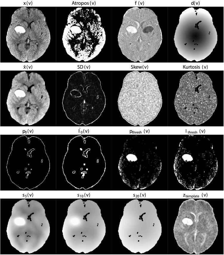
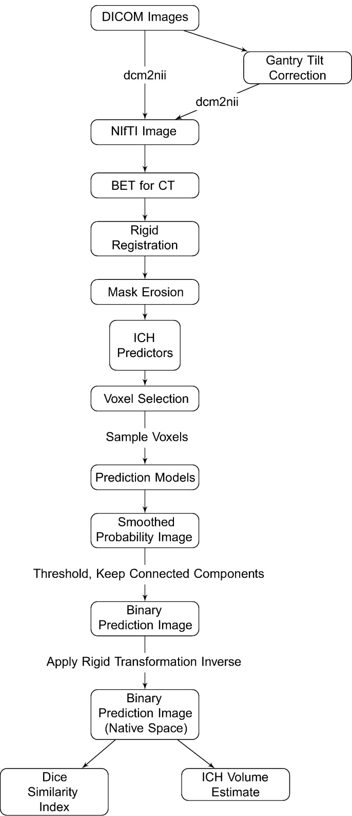
The Minimally Invasive Surgery plus rt-PA in ICH Evacuation (MISTIE) trial was a multi-site Phase II clinical trial that tested the safety of hemorrhage removal using recombinant-tissue plasminogen activator (rt-PA). For this analysis, we use 112 baseline CT scans from patients enrolled in the MISTE trial, one CT scan per patient. ICH was manually segmented on these CT scans by expert readers. We derived a set of imaging predictors from each scan. Using 10 randomly-selected scans, we used a first-pass voxel selection procedure based on quantiles of a set of predictors and then built 4 models estimating the voxel-level probability of ICH. The models used were: 1) logistic regression, 2) logistic regression with a penalty on the model parameters using LASSO, 3) a generalized additive model (GAM) and 4) a random forest classifier. The remaining 102 scans were used for model validation.For each validation scan, the model predicted the probability of ICH at each voxel. These voxel-level probabilities were then thresholded to produce binary segmentations of the hemorrhage. These masks were compared to the manual segmentations using the Dice Similarity Index (DSI) and the correlation of hemorrhage volume of between the two segmentations. We tested equality of median DSI using the Kruskal-Wallis test across the 4 models. We tested equality of the median DSI from sets of 2 models using a Wilcoxon signed-rank test.
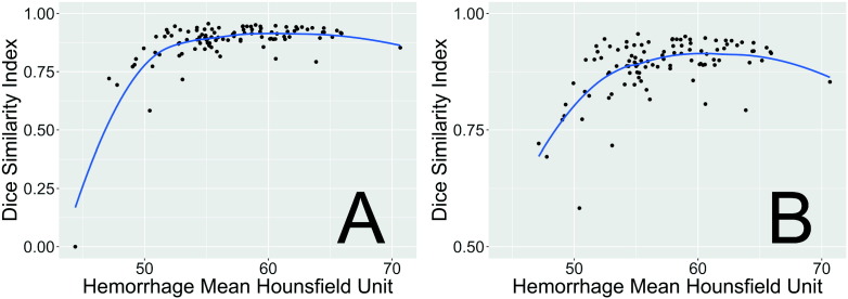
All results presented are for the 102 scans in the validation set. The median DSI for each model was: 0.89 (logistic), 0.885 (LASSO), 0.88 (GAM), and 0.899 (random forest). Using the random forest results in a slightly higher median DSI compared to the other models. After Bonferroni correction, the hypothesis of equality of median DSI was rejected only when comparing the random forest DSI to the DSI from the logistic ( < 0.001), LASSO ( < 0.001), or GAM ( < 0.001) models. In practical terms the difference between the random forest and the logistic regression is quite small. The correlation (95% CI) between the volume from manual segmentation and the predicted volume was 0.93 (0.9,0.95) for the random forest model. These results indicate that random forest approach can achieve accurate segmentation of ICH in a population of patients from a variety of imaging centers. We provide an R package (https://github.com/muschellij2/ichseg) and a Shiny R application online (http://johnmuschelli.com/ich_segment_all.html) for implementing and testing the proposed approach.
脑出血(ICH)是指血管破裂进入脑内区域,约占所有中风的10 - 15%。X射线计算机断层扫描(CT)主要用于评估这些出血的位置和体积。由专业阅片者使用面积测量法对CT扫描进行手动分割是体积估计的金标准,但耗时且阅片者内部和之间存在变异性。我们提出一种使用随机森林算法的全自动分割方法,该算法的特征从X射线计算机断层扫描(CT)中提取。
脑出血微创外科手术联合rt - PA清除术(MISTIE)试验是一项多中心II期临床试验,测试了使用重组组织型纤溶酶原激活剂(rt - PA)清除血肿的安全性。对于此分析,我们使用了MISTIE试验中入组患者的112份基线CT扫描,每位患者一份CT扫描。由专业阅片者在这些CT扫描上对脑出血进行手动分割。我们从每次扫描中得出一组影像预测指标。使用10份随机选择的扫描,我们基于一组预测指标的分位数采用首次体素选择程序,然后构建4个估计脑出血体素级概率的模型。所使用的模型为:1)逻辑回归,2)对模型参数使用LASSO进行惩罚的逻辑回归,3)广义相加模型(GAM),4)随机森林分类器。其余102份扫描用于模型验证。对于每份验证扫描,模型预测每个体素处脑出血的概率。然后对这些体素级概率进行阈值处理以生成血肿的二元分割。使用骰子相似性指数(DSI)以及两种分割之间血肿体积的相关性将这些掩码与手动分割进行比较。我们使用Kruskal - Wallis检验在4个模型之间测试中位数DSI的相等性。我们使用Wilcoxon符号秩检验在2个模型组之间测试中位数DSI的相等性。
呈现的所有结果均针对验证集中的102份扫描。每个模型的中位数DSI为:0.89(逻辑回归),0.885(LASSO),0.88(GAM)和0.899(随机森林)。与其他模型相比,使用随机森林得到的中位数DSI略高。经过Bonferroni校正后,仅在将随机森林DSI与逻辑回归(<0.001)、LASSO(<0.001)或GAM(<0.001)模型的DSI进行比较时,中位数DSI相等的假设被拒绝。实际上,随机森林与逻辑回归之间的差异非常小。随机森林模型中手动分割体积与预测体积之间的相关性(95%CI)为0.93(0.9,0.95)。这些结果表明,随机森林方法可以在来自不同影像中心的患者群体中实现脑出血的准确分割。我们提供了一个R包(https://github.com/muschellij2/ichseg)和一个在线Shiny R应用程序(http://johnmuschelli.com/ich_segment_all.html)来实施和测试所提出的方法。