• 文献检索
  • 文档翻译
  • 深度研究
  • 学术资讯
  • Suppr Zotero 插件Zotero 插件
  • 邀请有礼
  • 套餐&价格
  • 历史记录
应用&插件
Suppr Zotero 插件Zotero 插件浏览器插件Mac 客户端Windows 客户端微信小程序
定价
高级版会员购买积分包购买API积分包
服务
文献检索文档翻译深度研究API 文档MCP 服务
关于我们
关于 Suppr公司介绍联系我们用户协议隐私条款
关注我们

Suppr 超能文献

核心技术专利:CN118964589B侵权必究
粤ICP备2023148730 号-1Suppr @ 2026

文献检索

告别复杂PubMed语法,用中文像聊天一样搜索,搜遍4000万医学文献。AI智能推荐,让科研检索更轻松。

立即免费搜索

文件翻译

保留排版,准确专业,支持PDF/Word/PPT等文件格式,支持 12+语言互译。

免费翻译文档

深度研究

AI帮你快速写综述,25分钟生成高质量综述,智能提取关键信息,辅助科研写作。

立即免费体验

使用高分辨率 7T 时间飞跃血管造影术对人脑的脑膜动脉血管进行活体成像。

Imaging of the pial arterial vasculature of the human brain in vivo using high-resolution 7T time-of-flight angiography.

机构信息

Athinoula A. Martinos Center for Biomedical Imaging, Massachusetts General Hospital, Charlestown, United States.

Department of Radiology, Harvard Medical School, Boston, United States.

出版信息

Elife. 2022 Apr 29;11:e71186. doi: 10.7554/eLife.71186.

DOI:10.7554/eLife.71186
PMID:35486089
原文链接:https://pmc.ncbi.nlm.nih.gov/articles/PMC9150892/
Abstract

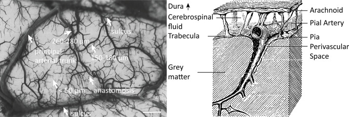
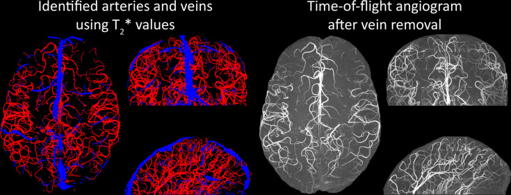
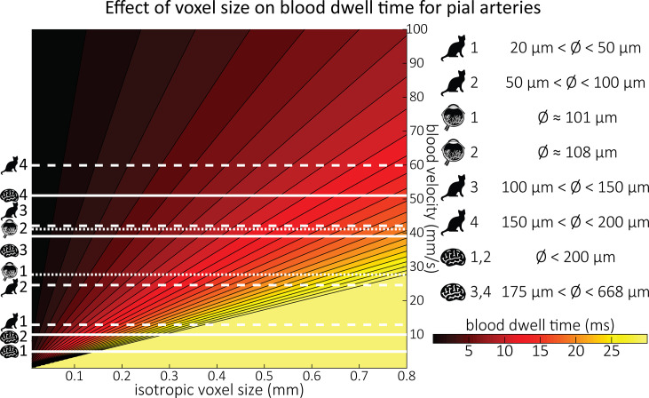
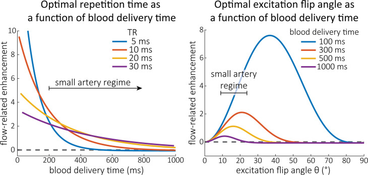
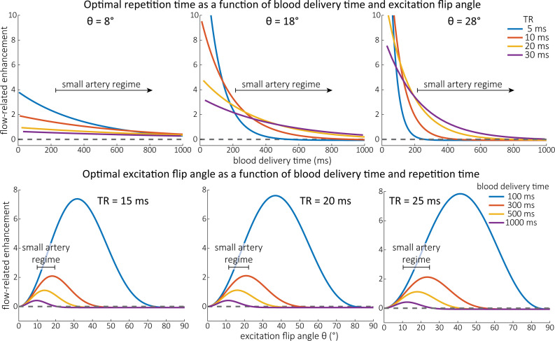
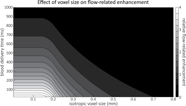
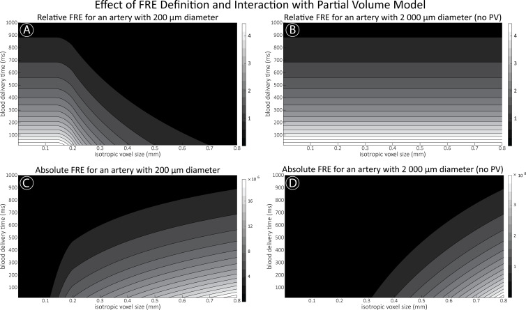
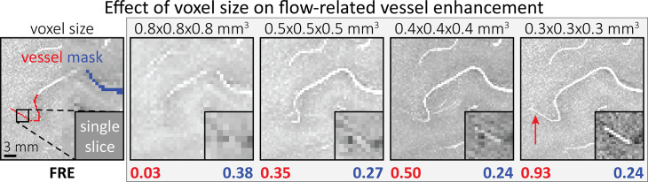
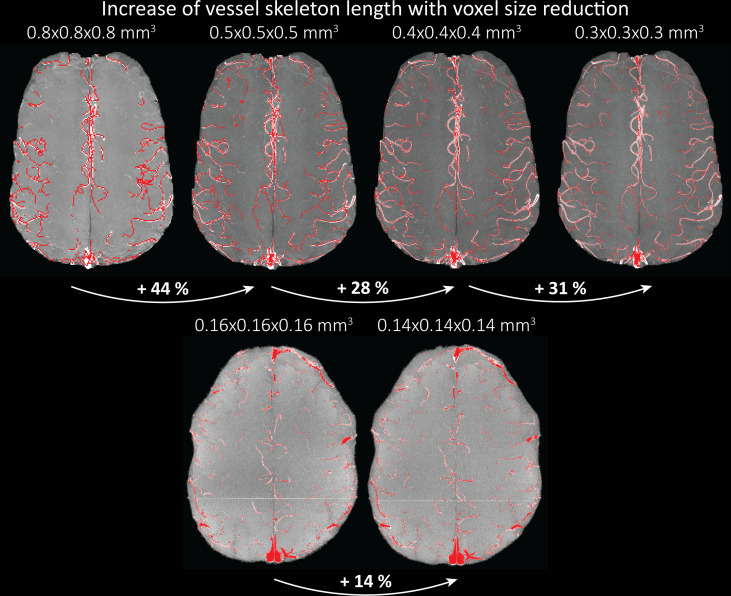
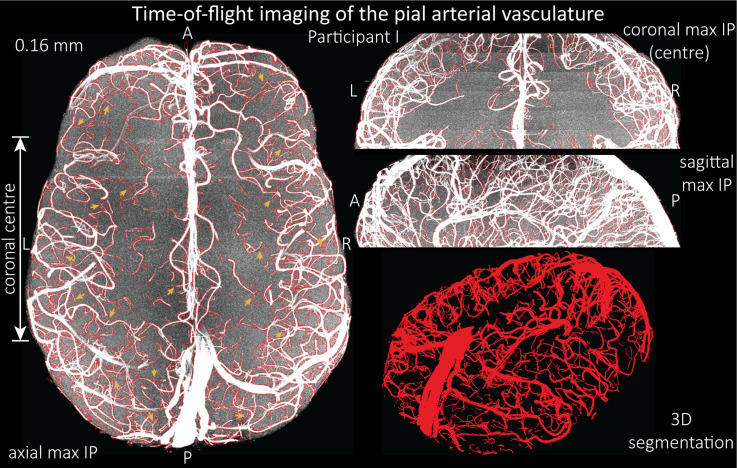
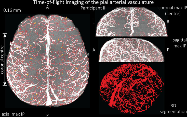
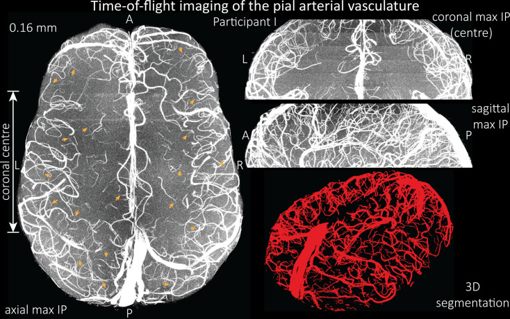
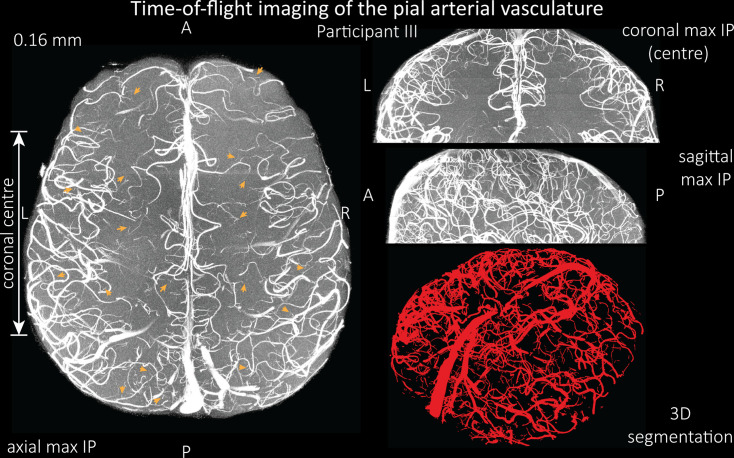
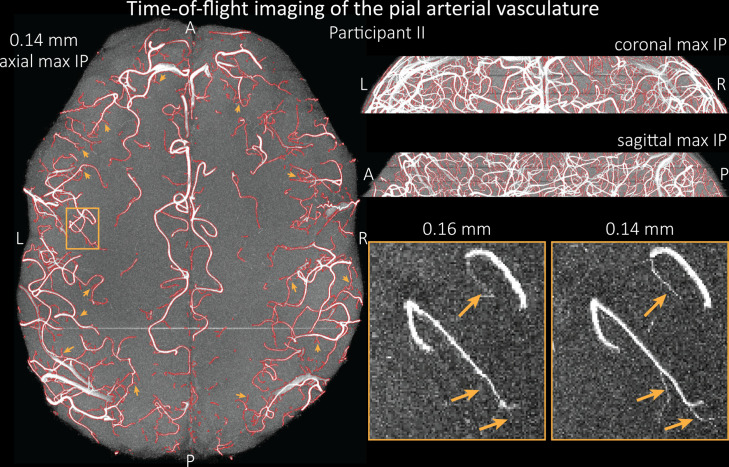
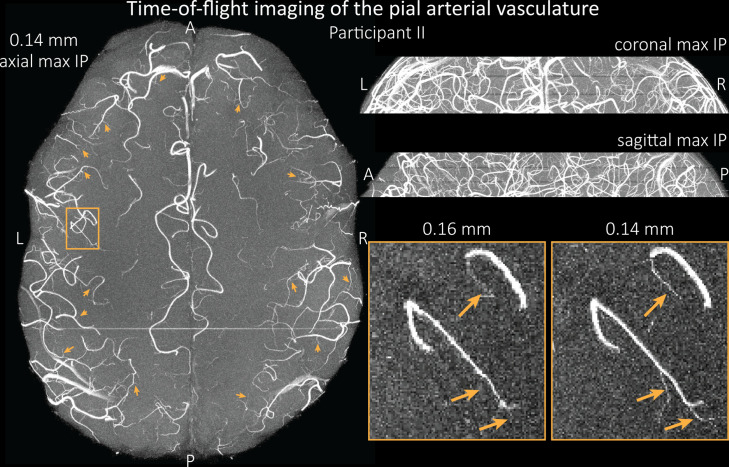
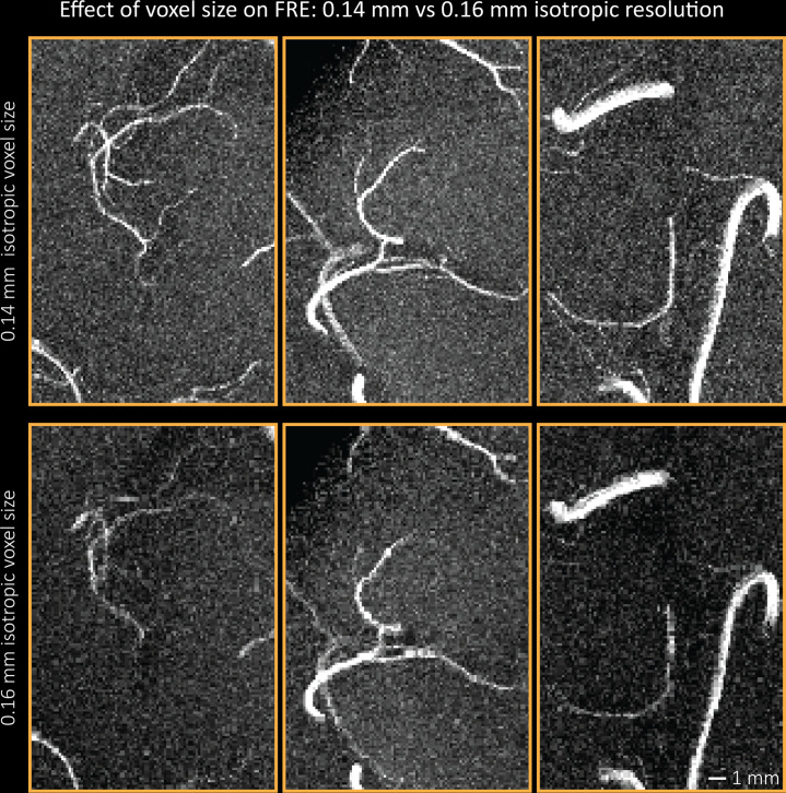
The pial arterial vasculature of the human brain is the only blood supply to the neocortex, but quantitative data on the morphology and topology of these mesoscopic arteries (diameter 50-300 µm) remains scarce. Because it is commonly assumed that blood flow velocities in these vessels are prohibitively slow, non-invasive time-of-flight magnetic resonance angiography (TOF-MRA)-which is well suited to high 3D imaging resolutions-has not been applied to imaging the pial arteries. Here, we provide a theoretical framework that outlines how TOF-MRA can visualize small pial arteries in vivo, by employing extremely small voxels at the size of individual vessels. We then provide evidence for this theory by imaging the pial arteries at 140 µm isotropic resolution using a 7 Tesla (T) magnetic resonance imaging (MRI) scanner and prospective motion correction, and show that pial arteries one voxel width in diameter can be detected. We conclude that imaging pial arteries is not limited by slow blood flow, but instead by achievable image resolution. This study represents the first targeted, comprehensive account of imaging pial arteries in vivo in the human brain. This ultra-high-resolution angiography will enable the characterization of pial vascular anatomy across the brain to investigate patterns of blood supply and relationships between vascular and functional architecture.

摘要

人脑的脑膜动脉血管是新皮质的唯一血液供应,但这些中观动脉(直径 50-300μm)的形态和拓扑结构的定量数据仍然很少。由于通常假设这些血管中的血流速度非常缓慢,因此不适合进行非侵入性的时间飞跃磁共振血管造影术(TOF-MRA)-这种技术非常适合高 3D 成像分辨率-尚未应用于脑膜动脉成像。在这里,我们提供了一个理论框架,概述了如何通过使用单个血管大小的极小体素来可视化体内的小脑膜动脉。然后,我们通过在 7 特斯拉(T)磁共振成像(MRI)扫描仪上使用 140μm 各向同性分辨率和前瞻性运动校正来对脑膜动脉进行成像,并提供了这一理论的证据,表明可以检测到直径为一个体素宽度的脑膜动脉。我们的结论是,脑膜动脉的成像不受血流缓慢的限制,而是受可实现的图像分辨率的限制。这项研究代表了首次在人类大脑中针对脑膜动脉进行体内成像的全面综合描述。这种超高分辨率血管造影术将能够描述整个大脑的脑膜血管解剖结构,以研究血液供应模式和血管与功能结构之间的关系。

https://cdn.ncbi.nlm.nih.gov/pmc/blobs/6a61/9150892/30ff1f6cda39/elife-71186-fig9.jpg
https://cdn.ncbi.nlm.nih.gov/pmc/blobs/6a61/9150892/459a47f48f0a/elife-71186-fig1.jpg
https://cdn.ncbi.nlm.nih.gov/pmc/blobs/6a61/9150892/a88964cd1d15/elife-71186-fig2.jpg
https://cdn.ncbi.nlm.nih.gov/pmc/blobs/6a61/9150892/55d9661d6353/elife-71186-fig3.jpg
https://cdn.ncbi.nlm.nih.gov/pmc/blobs/6a61/9150892/87926fe36a49/elife-71186-fig3-figsupp1.jpg
https://cdn.ncbi.nlm.nih.gov/pmc/blobs/6a61/9150892/804c2d2a2a97/elife-71186-fig4.jpg
https://cdn.ncbi.nlm.nih.gov/pmc/blobs/6a61/9150892/545dc4508d00/elife-71186-fig4-figsupp1.jpg
https://cdn.ncbi.nlm.nih.gov/pmc/blobs/6a61/9150892/9d6ccb932fe7/elife-71186-fig4-figsupp2.jpg
https://cdn.ncbi.nlm.nih.gov/pmc/blobs/6a61/9150892/2c23e203dd28/elife-71186-fig5.jpg
https://cdn.ncbi.nlm.nih.gov/pmc/blobs/6a61/9150892/3c0e14be55f2/elife-71186-fig5-figsupp1.jpg
https://cdn.ncbi.nlm.nih.gov/pmc/blobs/6a61/9150892/55427ed6674c/elife-71186-fig6.jpg
https://cdn.ncbi.nlm.nih.gov/pmc/blobs/6a61/9150892/f104519d9b37/elife-71186-fig6-figsupp1.jpg
https://cdn.ncbi.nlm.nih.gov/pmc/blobs/6a61/9150892/3f56fa1c7767/elife-71186-fig6-figsupp2.jpg
https://cdn.ncbi.nlm.nih.gov/pmc/blobs/6a61/9150892/ff1ed0099230/elife-71186-fig6-figsupp3.jpg
https://cdn.ncbi.nlm.nih.gov/pmc/blobs/6a61/9150892/147318b16354/elife-71186-fig7.jpg
https://cdn.ncbi.nlm.nih.gov/pmc/blobs/6a61/9150892/f6b7e53c9add/elife-71186-fig7-figsupp1.jpg
https://cdn.ncbi.nlm.nih.gov/pmc/blobs/6a61/9150892/d1c247258e13/elife-71186-fig7-figsupp2.jpg
https://cdn.ncbi.nlm.nih.gov/pmc/blobs/6a61/9150892/57d6deb2f89b/elife-71186-fig7-figsupp3.jpg
https://cdn.ncbi.nlm.nih.gov/pmc/blobs/6a61/9150892/2e3382a060b6/elife-71186-fig8.jpg
https://cdn.ncbi.nlm.nih.gov/pmc/blobs/6a61/9150892/2a3af834c99c/elife-71186-fig8-figsupp1.jpg
https://cdn.ncbi.nlm.nih.gov/pmc/blobs/6a61/9150892/30ff1f6cda39/elife-71186-fig9.jpg

相似文献

1
Imaging of the pial arterial vasculature of the human brain in vivo using high-resolution 7T time-of-flight angiography.使用高分辨率 7T 时间飞跃血管造影术对人脑的脑膜动脉血管进行活体成像。
Elife. 2022 Apr 29;11:e71186. doi: 10.7554/eLife.71186.
2
Prospective motion correction enables highest resolution time-of-flight angiography at 7T.前瞻性运动校正可实现 7T 时最高分辨率的时间飞跃血管造影。
Magn Reson Med. 2018 Jul;80(1):248-258. doi: 10.1002/mrm.27033. Epub 2017 Dec 11.
3
Ultra high field TOF-MRA: A method to visualize small cerebral vessels. 7T TOF-MRA sequence parameters on different MRI scanners - Literature review.超高场 TOF-MRA:一种可视化小脑血管的方法。不同 MRI 扫描仪上的 7T TOF-MRA 序列参数——文献综述。
Neurol Neurochir Pol. 2017 Sep-Oct;51(5):411-418. doi: 10.1016/j.pjnns.2017.06.011. Epub 2017 Jul 8.
4
3 T contrast-enhanced magnetic resonance angiography for evaluation of the intracranial arteries: comparison with time-of-flight magnetic resonance angiography and multislice computed tomography angiography.3T对比增强磁共振血管造影术用于评估颅内动脉:与时间飞跃磁共振血管造影术及多层螺旋CT血管造影术的比较
Invest Radiol. 2006 Nov;41(11):799-805. doi: 10.1097/01.rli.0000242835.00032.f5.
5
High resolution time-of-flight MR-angiography at 7 T exploiting VERSE saturation, compressed sensing and segmentation.7T 下利用 VERSE 饱和、压缩感知和分割进行高分辨率时间飞跃磁共振血管造影。
Magn Reson Imaging. 2019 Nov;63:193-204. doi: 10.1016/j.mri.2019.08.014. Epub 2019 Aug 18.
6
Highly accelerated time-of-flight magnetic resonance angiography using spiral imaging improves conspicuity of intracranial arterial branches while reducing scan time.采用螺旋成像的高速时间飞跃磁共振血管造影可在减少扫描时间的同时提高颅内动脉分支的显影效果。
Eur Radiol. 2020 Feb;30(2):855-865. doi: 10.1007/s00330-019-06442-y. Epub 2019 Oct 29.
7
Added diagnostic values of three-dimensional high-resolution proton density-weighted magnetic resonance imaging for unruptured intracranial aneurysms in the circle-of-Willis: Comparison with time-of-flight magnetic resonance angiography.三维高分辨质子密度加权磁共振成像对颅内未破裂动脉瘤的诊断价值:与时间飞跃磁共振血管造影的比较。
PLoS One. 2020 Dec 3;15(12):e0243235. doi: 10.1371/journal.pone.0243235. eCollection 2020.
8
Evaluation of the applicability of territorial arterial spin labeling in meningiomas for presurgical assessments compared with 3-dimensional time-of-flight magnetic resonance angiography.评价基于体素的动脉自旋标记在脑膜瘤术前评估中的适用性,与 3 维时间飞跃磁共振血管成像相比。
Eur Radiol. 2017 Oct;27(10):4072-4081. doi: 10.1007/s00330-017-4760-9. Epub 2017 Mar 16.
9
Digital reconstruction and morphometric analysis of human brain arterial vasculature from magnetic resonance angiography.基于磁共振血管成像的人脑动脉血管的数字重建与形态计量分析。
Neuroimage. 2013 Nov 15;82:170-81. doi: 10.1016/j.neuroimage.2013.05.089. Epub 2013 May 28.
10
Clinical vascular imaging in the brain at 7T.7T 下脑部的临床血管成像。
Neuroimage. 2018 Mar;168:452-458. doi: 10.1016/j.neuroimage.2016.11.044. Epub 2016 Nov 18.

引用本文的文献

1
Macrovascular contributions to resting-state fMRI signals: A comparison between EPI and bSSFP at 9.4 Tesla.大血管对静息态功能磁共振成像信号的贡献:9.4特斯拉下回波平面成像(EPI)与稳态自由进动序列(bSSFP)的比较
Imaging Neurosci (Camb). 2025 Jan 7;3. doi: 10.1162/imag_a_00435. eCollection 2025.
2
Characterisation of laminar and vascular spatiotemporal dynamics of CBV and BOLD signals using VASO and ME-GRE at 7T in humans.在7T磁场下,使用动脉自旋标记(VASO)和多回波梯度回波(ME-GRE)对人类脑血容量(CBV)和血氧水平依赖(BOLD)信号的层流和血管时空动力学进行表征。
Imaging Neurosci (Camb). 2024 Aug 13;2. doi: 10.1162/imag_a_00263. eCollection 2024.
3

本文引用的文献

1
TAPAS: An Open-Source Software Package for Translational Neuromodeling and Computational Psychiatry.TAPAS:一个用于转化神经建模和计算精神病学的开源软件包。
Front Psychiatry. 2021 Jun 2;12:680811. doi: 10.3389/fpsyt.2021.680811. eCollection 2021.
2
Comprehensive ultrahigh resolution whole brain in vivo MRI dataset as a human phantom.作为一个人体模型的全面超高分辨率全脑活体 MRI 数据集。
Sci Data. 2021 May 25;8(1):138. doi: 10.1038/s41597-021-00923-w.
3
Improved susceptibility weighted imaging at ultra-high field using bipolar multi-echo acquisition and optimized image processing: CLEAR-SWI.
Assessing Arterial Patterns in the Motor Cortex With 7 Tesla Magnetic Resonance Imaging and Vessel Distance Mapping.
利用7特斯拉磁共振成像和血管距离映射评估运动皮层的动脉模式。
Hum Brain Mapp. 2025 Aug 1;46(11):e70311. doi: 10.1002/hbm.70311.
4
Non-contrast enhanced visualization of the equine foot vasculature in a cadaver model using time-of-flight sequence.在尸体模型中使用飞行时间序列对马足部血管系统进行非增强可视化。
Front Vet Sci. 2025 Jul 18;12:1585940. doi: 10.3389/fvets.2025.1585940. eCollection 2025.
5
Mapping cerebral blood perfusion and its links to multi-scale brain organization across the human lifespan.绘制全人类生命周期内的脑血流灌注及其与多尺度脑组织的联系。
PLoS Biol. 2025 Jul 29;23(7):e3003277. doi: 10.1371/journal.pbio.3003277. eCollection 2025 Jul.
6
Mapping vascular network architecture in primate brain using ferumoxytol-weighted laminar MRI.使用菲洛米星加权层流MRI绘制灵长类动物大脑中的血管网络结构。
Elife. 2025 Jun 11;13:RP99940. doi: 10.7554/eLife.99940.
7
Using PINS pulses to saturate inflow effects on fMRI data at 3 and 7 T.使用PINS脉冲在3T和7T场强下饱和流入效应以获取功能磁共振成像数据。
Magn Reson Med. 2025 Oct;94(4):1635-1643. doi: 10.1002/mrm.30584. Epub 2025 May 20.
8
Characterizing positive and negative quantitative susceptibility values in the cortex following mild traumatic brain injury: a depth- and curvature-based study.轻度创伤性脑损伤后皮质中正负定量磁化率值的特征:一项基于深度和曲率的研究。
Cereb Cortex. 2025 Mar 6;35(3). doi: 10.1093/cercor/bhaf059.
9
Deep learning for 3D vascular segmentation in hierarchical phase contrast tomography: a case study on kidney.深度学习在分层相衬断层摄影中的 3D 血管分割:以肾脏为例的研究
Sci Rep. 2024 Nov 8;14(1):27258. doi: 10.1038/s41598-024-77582-5.
10
Self-supervised learning for accurately modelling hierarchical evolutionary patterns of cerebrovasculature.基于自监督学习的脑循环系统进化模式精准建模。
Nat Commun. 2024 Oct 25;15(1):9235. doi: 10.1038/s41467-024-53550-5.
采用双极多回波采集和优化图像处理的超高场改进磁敏感加权成像:CLEAR-SWI。
Neuroimage. 2021 Aug 15;237:118175. doi: 10.1016/j.neuroimage.2021.118175. Epub 2021 May 15.
4
Velocity and Pulsatility Measures in the Perforating Arteries of the Basal Ganglia at 3T MRI in Reference to 7T MRI.3T磁共振成像(MRI)下基底节区穿支动脉的速度和搏动性测量:与7T MRI的对比研究
Front Neurosci. 2021 Apr 26;15:665480. doi: 10.3389/fnins.2021.665480. eCollection 2021.
5
BRAVE-NET: Fully Automated Arterial Brain Vessel Segmentation in Patients With Cerebrovascular Disease.BRAVE-NET:脑血管疾病患者的全自动脑动脉血管分割
Front Artif Intell. 2020 Sep 25;3:552258. doi: 10.3389/frai.2020.552258. eCollection 2020.
6
DeepVesselNet: Vessel Segmentation, Centerline Prediction, and Bifurcation Detection in 3-D Angiographic Volumes.深度血管网络:三维血管造影体积中的血管分割、中心线预测和分叉检测
Front Neurosci. 2020 Dec 8;14:592352. doi: 10.3389/fnins.2020.592352. eCollection 2020.
7
Origin of the Time Lag Phenomenon and the Global Signal in Resting-State fMRI.静息态功能磁共振成像中时间滞后现象和全局信号的起源
Front Neurosci. 2020 Oct 29;14:596084. doi: 10.3389/fnins.2020.596084. eCollection 2020.
8
Cerebral artery segmentation based on magnetization-prepared two rapid acquisition gradient echo multi-contrast images in 7 Tesla magnetic resonance imaging.基于 7T 磁共振成像中磁化准备双快速获取梯度回波多对比度图像的脑动脉分割。
Neuroimage. 2020 Nov 15;222:117259. doi: 10.1016/j.neuroimage.2020.117259. Epub 2020 Aug 13.
9
Ultra-slow Oscillations in fMRI and Resting-State Connectivity: Neuronal and Vascular Contributions and Technical Confounds.功能磁共振成像和静息态连通性中的超慢震荡:神经元和血管的贡献及技术混淆。
Neuron. 2020 Sep 9;107(5):782-804. doi: 10.1016/j.neuron.2020.07.020. Epub 2020 Aug 12.
10
Multiscale modeling of human cerebrovasculature: A hybrid approach using image-based geometry and a mathematical algorithm.人体脑血管的多尺度建模:基于图像的几何形状和数学算法的混合方法。
PLoS Comput Biol. 2020 Jun 22;16(6):e1007943. doi: 10.1371/journal.pcbi.1007943. eCollection 2020 Jun.