Shi Mengjie, Zhao Tianrui, West Simeon J, Desjardins Adrien E, Vercauteren Tom, Xia Wenfeng
School of Biomedical Engineering and Imaging Sciences, King's College London, London SE1 7EH, United Kingdom.
Department of Anaesthesia, University College Hospital, London NW1 2BU, United Kingdom.
Photoacoustics. 2022 Apr 7;26:100351. doi: 10.1016/j.pacs.2022.100351. eCollection 2022 Jun.
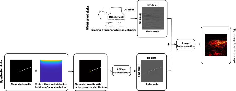
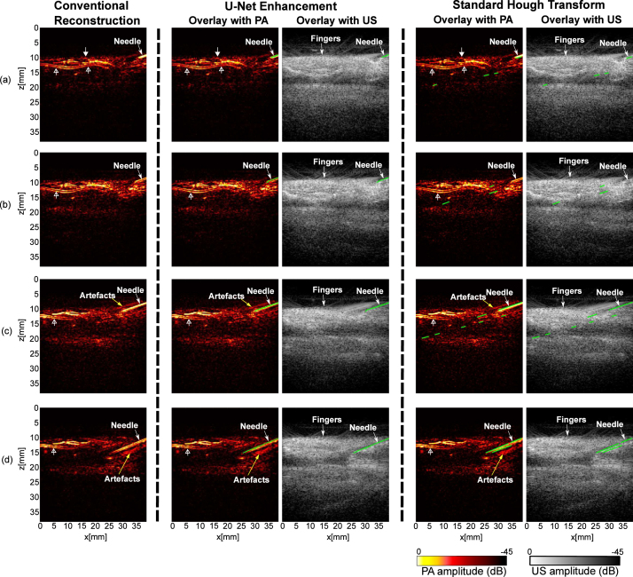
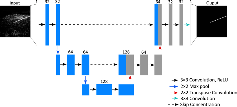
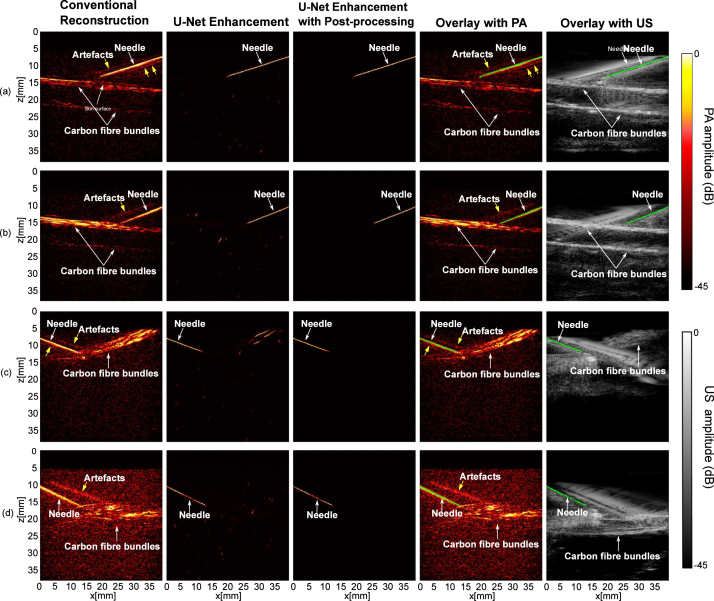
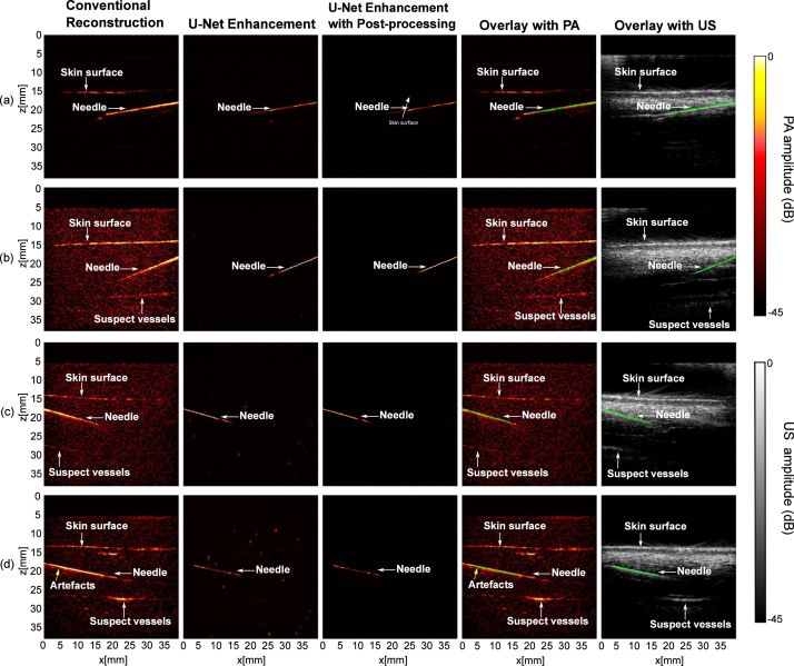
Photoacoustic imaging has shown great potential for guiding minimally invasive procedures by accurate identification of critical tissue targets and invasive medical devices (such as metallic needles). The use of light emitting diodes (LEDs) as the excitation light sources accelerates its clinical translation owing to its high affordability and portability. However, needle visibility in LED-based photoacoustic imaging is compromised primarily due to its low optical fluence. In this work, we propose a deep learning framework based on U-Net to improve the visibility of clinical metallic needles with a LED-based photoacoustic and ultrasound imaging system. To address the complexity of capturing ground truth for real data and the poor realism of purely simulated data, this framework included the generation of semi-synthetic training datasets combining both simulated data to represent features from the needles and measurements for tissue background. Evaluation of the trained neural network was performed with needle insertions into blood-vessel-mimicking phantoms, pork joint tissue and measurements on human volunteers. This deep learning-based framework substantially improved the needle visibility in photoacoustic imaging compared to conventional reconstruction by suppressing background noise and image artefacts, achieving 5.8 and 4.5 times improvements in terms of signal-to-noise ratio and the modified Hausdorff distance, respectively. Thus, the proposed framework could be helpful for reducing complications during percutaneous needle insertions by accurate identification of clinical needles in photoacoustic imaging.
光声成像在通过精确识别关键组织靶点和侵入性医疗设备(如金属针)来指导微创手术方面已显示出巨大潜力。使用发光二极管(LED)作为激发光源,因其高性价比和便携性,加速了其临床转化。然而,基于LED的光声成像中针的可见性主要因其低光通量而受到影响。在这项工作中,我们提出了一种基于U-Net的深度学习框架,以提高基于LED的光声和超声成像系统中临床金属针的可见性。为了解决获取真实数据的地面真值的复杂性以及纯模拟数据的逼真度差的问题,该框架包括生成半合成训练数据集,该数据集结合了表示针特征的模拟数据和组织背景的测量数据。通过将针插入血管模拟体模、猪肉关节组织以及对人类志愿者进行测量,对训练后的神经网络进行了评估。与传统重建相比,这种基于深度学习的框架通过抑制背景噪声和图像伪影,显著提高了光声成像中针的可见性,在信噪比和修正的豪斯多夫距离方面分别提高了5.8倍和4.5倍。因此,所提出的框架有助于通过在光声成像中准确识别临床针来减少经皮针插入过程中的并发症。