Ranasinghe Meenakshi, Arifuzzaman Md, Rajamanthrilage Apeksha C, Willoughby W R, Dickey Ashley, McMillen Colin, Kolis Joseph W, Bolding Mark, Anker Jeffrey N
Department of Chemistry, Center for Optical Materials Engineering and Technology (COMSET), Clemson University Clemson SC USA
Department of Radiology, University of Alabama at Birmingham School of Medicine Birmingham AL USA.
RSC Adv. 2021 Sep 24;11(50):31717-31726. doi: 10.1039/d1ra05451a. eCollection 2021 Sep 21.
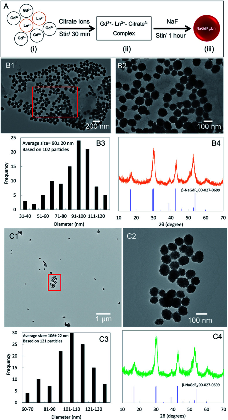
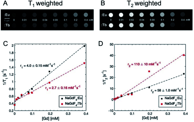
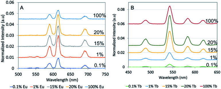
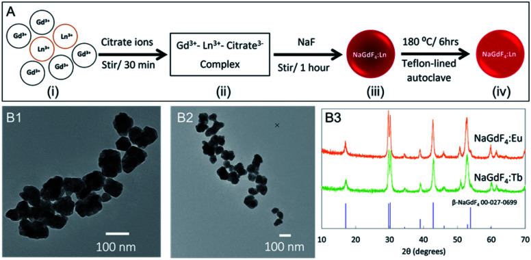
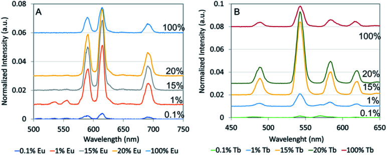
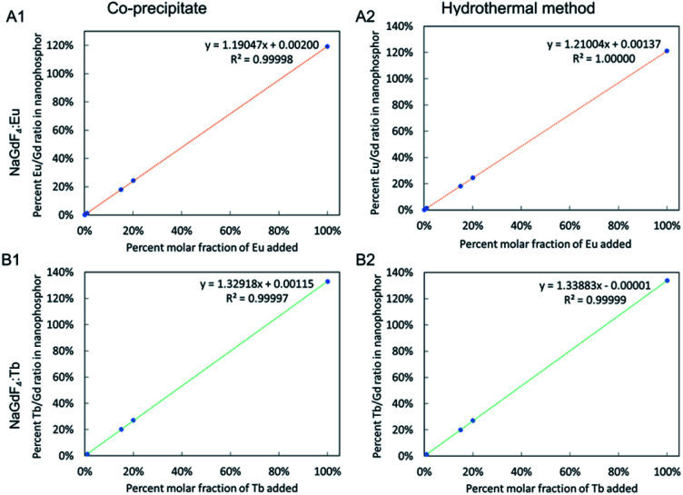
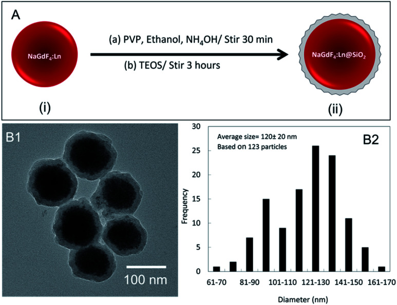
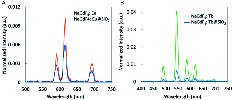
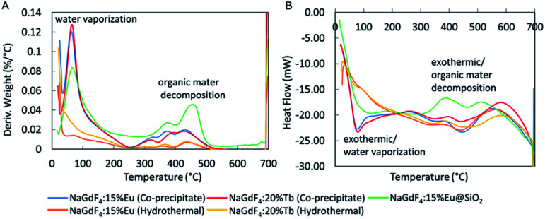
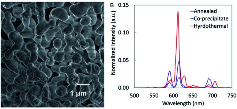
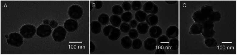
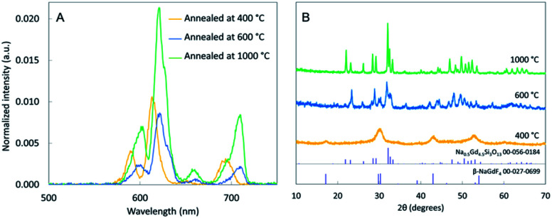
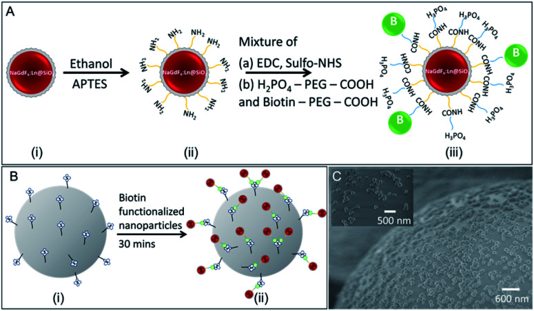
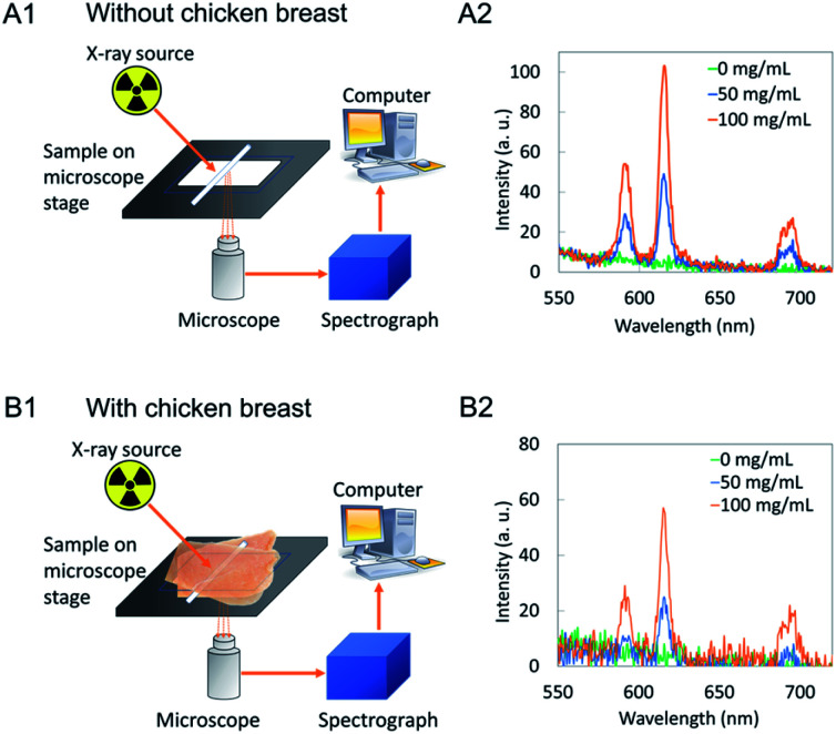
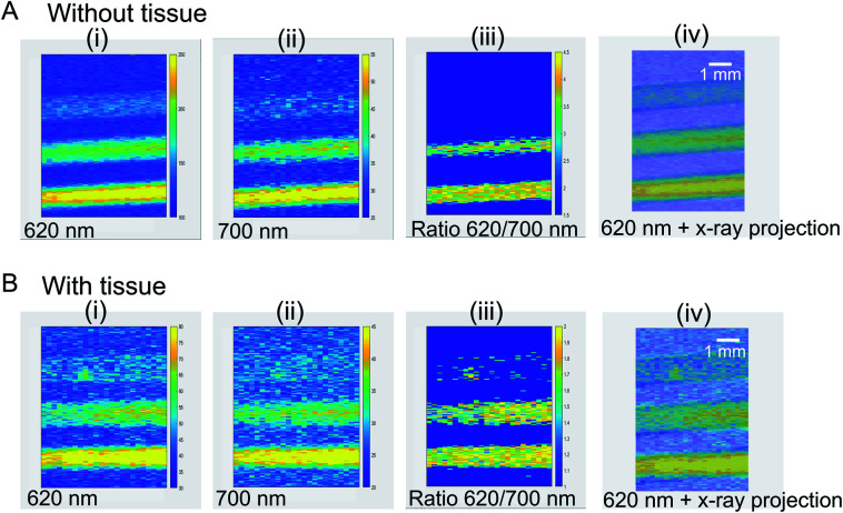
X-ray excited optical luminescence from nanophosphors can be used to selectively generate light in tissue for imaging and stimulating light-responsive materials and cells. Herein, we synthesized X-ray scintillating NaGdF:Eu and Tb nanophosphors co-precipitate and hydrothermal methods, encapsulated with silica, functionalized with biotin, and characterized by X-ray excited optical luminescence spectroscopy and imaging. The nanophosphors synthesized by co-precipitate method were ∼90 and ∼106 nm in diameter, respectively, with hydrothermally synthesized particles showing the highest luminescence intensity. More importantly, we investigated the effect of thermal annealing/calcination on the X-ray excited luminescence spectra and intensity. At above 1000 °C, the luminescence intensity increased, but particles fused together. Coating with a 15 nm thick silica shell prevented particle fusion and enabled silane-based chemical functionalization, although luminescence decreased largely due to the increased mass of non-luminescent material. We observed an increase in luminesce intensity with temperature until at 400 °C. At above 600 °C, NaGdF:Eu@SiO converts to NaGdSiO:Eu, an X-ray scintillator brighter than annealed NPs at 400 °C and dimmer than NPs synthesized using the hydrothermal method. The particles generate light through tissue and can be selectively excited using a focused X-ray source for imaging and light generation applications. The particles also act as MRI contrast agents for multi-modal localization.
纳米磷光体的X射线激发光学发光可用于在组织中选择性地产生光,以进行成像以及刺激光响应材料和细胞。在此,我们通过共沉淀法和水热法合成了X射线闪烁的NaGdF:Eu和Tb纳米磷光体,用二氧化硅包裹,用生物素功能化,并通过X射线激发光学发光光谱和成像进行表征。通过共沉淀法合成的纳米磷光体直径分别约为90和106 nm,水热合成的颗粒显示出最高的发光强度。更重要的是,我们研究了热退火/煅烧对X射线激发发光光谱和强度的影响。在1000℃以上,发光强度增加,但颗粒融合在一起。用15 nm厚的二氧化硅壳涂层可防止颗粒融合,并实现基于硅烷的化学功能化,尽管由于非发光材料质量增加,发光强度大幅下降。我们观察到发光强度随温度升高,直到400℃。在600℃以上,NaGdF:Eu@SiO转变为NaGdSiO:Eu,一种X射线闪烁体,在400℃时比退火的纳米颗粒亮,但比用水热法合成的纳米颗粒暗。这些颗粒可透过组织发光,并可使用聚焦X射线源进行选择性激发,用于成像和光生成应用。这些颗粒还可作为多模态定位的MRI造影剂。