Department of Ophthalmology, Chang Gung Memorial Hospital, Linkou Medical Center, Taoyuan, Taiwan.
College of Medicine, Chang Gung University, Taoyuan, Taiwan.
Invest Ophthalmol Vis Sci. 2022 May 2;63(5):5. doi: 10.1167/iovs.63.5.5.
To compare the manifestations of photoreceptors (PRs) in three hereditary optic neuropathies affected by primary mitochondrial dysfunction and discuss whether the retinal ganglion cells (RGCs) or the PRs are preferentially affected.
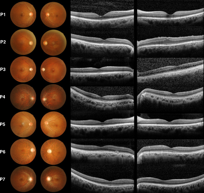
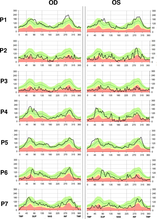
A retrospective analysis of patients with genetically confirmed diagnoses of optic neuropathies associated with mitochondrial dysfunction was performed. This cohort included Leber's hereditary optic neuropathy (LHON), autosomal dominant optic atrophy type 1 (OPA1), and optic atrophy type 13 (OPA13). Patient chart evaluations included clinical characteristics, best-corrected visual acuity (BCVA), fundus photography, spectral-domain optical coherence tomography (SD-OCT), electroretinogram (ERG), and visual evoked potential data.
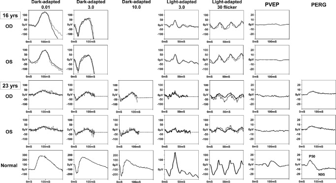
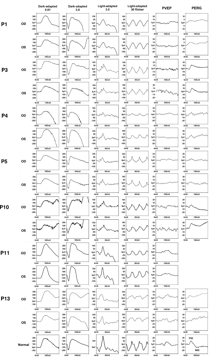
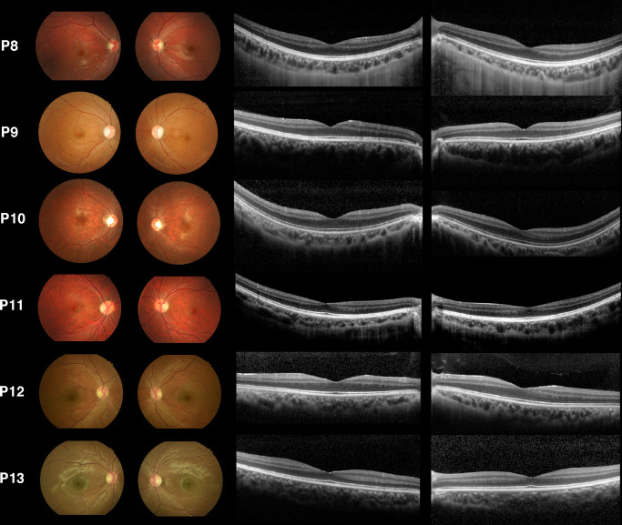
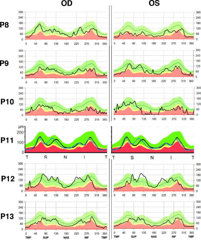
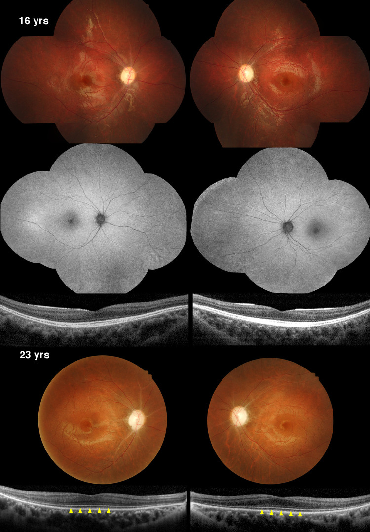
This analysis included seven patients with LHON, six with OPA1, and one with OPA13 from a tertiary medical center. Thirteen of the 14 individuals were male. The average BCVA at diagnosis was 20/285 and 20/500 in the right and left eyes, respectively. Five of the seven patients with LHON, and three of the six patients with OPA1 also showed a mild amplitude reduction or delayed latency on light-adapted ERG and 30-Hz flicker responses; however, SD-OCT imaging did not show correlated PR abnormalities. Notably, a 7-year follow-up of a patient with OPA13 revealed degeneration of RGCs prior to the degeneration of PRs. Follow-up data also demonstrated continuous loss of cone outer segment tips on SD-OCT imaging.
RGCs are, in general, affected by mitochondrial dysfunction, whereas variable PR dysfunction exists in patients with LHON and OPA1, especially with respect to the cone responses. Involvement of PRs is particularly evident in OPA13 after RGC degenerations.
比较三种由原发性线粒体功能障碍引起的遗传性视神经病变中光感受器(PRs)的表现,并探讨是视网膜神经节细胞(RGCs)还是 PRs 优先受到影响。
对经基因确诊的与线粒体功能障碍相关的视神经病变患者进行回顾性分析。该队列包括莱伯遗传性视神经病变(LHON)、常染色体显性视神经萎缩 1 型(OPA1)和视神经萎缩 13 型(OPA13)。患者图表评估包括临床特征、最佳矫正视力(BCVA)、眼底照相、谱域光相干断层扫描(SD-OCT)、视网膜电图(ERG)和视觉诱发电位数据。
这项分析包括来自三级医疗中心的 7 例 LHON 患者、6 例 OPA1 患者和 1 例 OPA13 患者。14 个人中有 13 个是男性。诊断时右眼平均 BCVA 为 20/285,左眼为 20/500。7 例 LHON 患者中的 5 例和 6 例 OPA1 患者中的 3 例,光适应 ERG 和 30-Hz 闪烁反应也表现出轻度幅度降低或潜伏期延迟;然而,SD-OCT 成像并未显示相关的 PR 异常。值得注意的是,OPA13 患者的 7 年随访显示,RGC 退化前 PR 已经退化。随访数据还表明,SD-OCT 成像上连续出现圆锥外节尖端丢失。
一般来说,RGC 受到线粒体功能障碍的影响,而 LHON 和 OPA1 患者的 PR 功能障碍不同,特别是在锥体细胞反应方面。OPA13 患者在 RGC 退化后,PR 明显受累。