Second Clinical Division, Peking University School and Hospital of Stomatology & National Clinical Research Center for Oral Diseases & National Engineering Laboratory for Digital and Material Technology of Stomatology & Beijing Key Laboratory of Digital Stomatology, 22 Zhongguancun South Avenue, Beijing, 100081, China.
Beijing Healya Technology Limited, Beijing, 100195, China.
Sci Rep. 2022 May 4;12(1):7217. doi: 10.1038/s41598-022-10065-7.
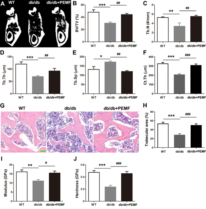
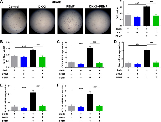
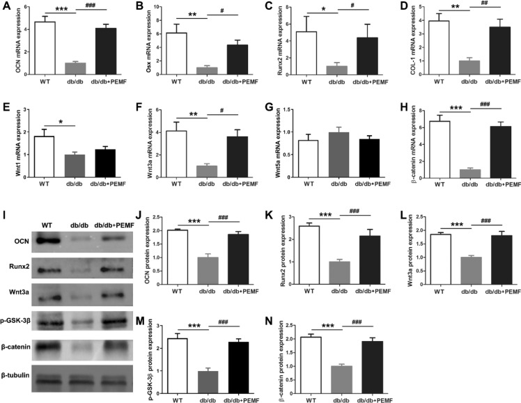
Type 2 diabetes mellitus (T2DM) patients have compromised mandibular bone architecture/quality, which markedly increase the risks of tooth loosening, tooth loss, and failure of dental implantation. However, it remains lacks effective and safe countermeasures against T2DM-related mandibular bone deterioration. Herein, we studied the effects of pulsed electromagnetic fields (PEMF) on mandibular bone microstructure/quality and relevant regulatory mechanisms in T2DM db/db mice. PEMF exposure (20 Gs, 15 Hz) for 12 weeks preserved trabecular bone architecture, increased cortical bone thickness, improved material properties and stimulated bone anabolism in mandibles of db/db mice. PEMF also upregulated the expression of canonical Wnt3a ligand (but not Wnt1 or Wnt5a) and its downstream β-catenin. PEMF improved the viability and differentiation of primary osteoblasts isolated from the db/db mouse mandible, and stimulated the specific activation of Wnt3a/β-catenin signaling. These positive effects of PEMF on mandibular osteoblasts of db/db mice were almost totally abolished after Wnt3a silencing in vitro, which were equivalent to the effects following blockade of canonical Wnt signaling using the broad-spectrum antagonist DKK1. Injection with Wnt3a siRNA abrogated the therapeutic effects of PEMF on mandibular bone quantity/quality and bone anabolism in db/db mice. Our study indicates that PEMF might become a non-invasive and safe treatment alternative resisting mandibular bone deterioration in T2DM patients, which is helpful for protecting teeth from loosening/loss and securing the dental implant stability.
2 型糖尿病(T2DM)患者的下颌骨结构/质量受损,这显著增加了牙齿松动、脱落和种植牙失败的风险。然而,目前仍然缺乏针对 T2DM 相关下颌骨恶化的有效和安全的对策。在此,我们研究了脉冲电磁场(PEMF)对 T2DM db/db 小鼠下颌骨微观结构/质量的影响及其相关调节机制。PEMF 暴露(20 Gs,15 Hz)12 周可保持骨小梁结构,增加皮质骨厚度,改善下颌骨的材料性能并刺激骨合成代谢。PEMF 还上调了经典 Wnt3a 配体(但不是 Wnt1 或 Wnt5a)及其下游β-连环蛋白的表达。PEMF 改善了从 db/db 小鼠下颌骨分离的原代成骨细胞的活力和分化,并刺激了 Wnt3a/β-连环蛋白信号的特异性激活。体外沉默 Wnt3a 后,PEMF 对 db/db 小鼠下颌骨成骨细胞的这些积极作用几乎完全被消除,这与使用广谱拮抗剂 DKK1 阻断经典 Wnt 信号通路的效果相当。Wnt3a siRNA 的注射消除了 PEMF 对 db/db 小鼠下颌骨数量/质量和骨合成代谢的治疗作用。我们的研究表明,PEMF 可能成为一种非侵入性和安全的治疗选择,用于抵抗 T2DM 患者的下颌骨恶化,有助于防止牙齿松动/脱落,并确保种植牙的稳定性。