Jing Da, Li Feijiang, Jiang Maogang, Cai Jing, Wu Yan, Xie Kangning, Wu Xiaoming, Tang Chi, Liu Juan, Guo Wei, Shen Guanghao, Luo Erping
Department of Biomedical Engineering, Fourth Military Medical University, Xi'an, China.
PLoS One. 2013 Nov 14;8(11):e79377. doi: 10.1371/journal.pone.0079377. eCollection 2013.
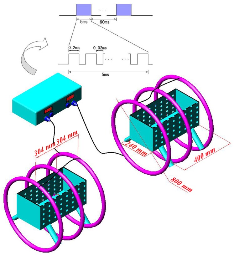
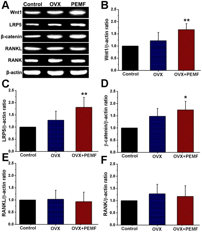
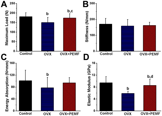
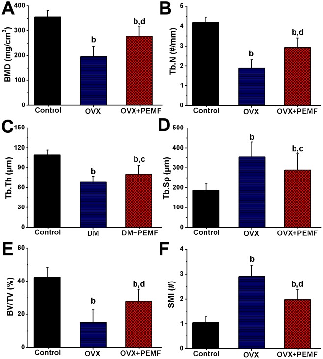
Growing evidence has demonstrated that pulsed electromagnetic field (PEMF), as an alternative noninvasive method, could promote remarkable in vivo and in vitro osteogenesis. However, the exact mechanism of PEMF on osteopenia/osteoporosis is still poorly understood, which further limits the extensive clinical application of PEMF. In the present study, the efficiency of PEMF on osteoporotic bone microarchitecture and bone quality together with its associated signaling pathway mechanisms was systematically investigated in ovariectomized (OVX) rats. Thirty rats were equally assigned to the Control, OVX and OVX+PEMF groups. The OVX+PEMF group was subjected to daily 8-hour PEMF exposure with 15 Hz, 2.4 mT (peak value). After 10 weeks, the OVX+PEMF group exhibited significantly improved bone mass and bone architecture, evidenced by increased BMD, Tb.N, Tb.Th and BV/TV, and suppressed Tb.Sp and SMI levels in the MicroCT analysis. Three-point bending test suggests that PEMF attenuated the biomechanical strength deterioration of the OVX rat femora, evidenced by increased maximum load and elastic modulus. RT-PCR analysis demonstrated that PEMF exposure significantly promoted the overall gene expressions of Wnt1, LRP5 and β-catenin in the canonical Wnt signaling, but did not exhibit obvious impact on either RANKL or RANK gene expressions. Together, our present findings highlight that PEMF attenuated OVX-induced deterioration of bone microarchitecture and strength in rats by promoting the activation of Wnt/LRP5/β-catenin signaling rather than by inhibiting RANKL-RANK signaling. This study enriches our basic knowledge to the osteogenetic activity of PEMF, and may lead to more efficient and scientific clinical application of PEMF in inhibiting osteopenia/osteoporosis.
越来越多的证据表明,脉冲电磁场(PEMF)作为一种替代性非侵入性方法,可在体内和体外显著促进成骨。然而,PEMF对骨质减少/骨质疏松的确切机制仍知之甚少,这进一步限制了PEMF在临床上的广泛应用。在本研究中,我们系统地研究了PEMF对去卵巢(OVX)大鼠骨质疏松性骨微结构和骨质量的影响及其相关信号通路机制。将30只大鼠平均分为对照组、OVX组和OVX+PEMF组。OVX+PEMF组每天接受8小时、频率15Hz、峰值2.4mT的PEMF暴露。10周后,MicroCT分析显示,OVX+PEMF组的骨量和骨结构显著改善,表现为骨密度、骨小梁数量、骨小梁厚度和骨体积分数增加,骨小梁间距和结构模型指数水平降低。三点弯曲试验表明,PEMF减轻了OVX大鼠股骨的生物力学强度恶化,表现为最大载荷和弹性模量增加。RT-PCR分析表明,PEMF暴露显著促进了经典Wnt信号通路中Wnt1、LRP5和β-连环蛋白的整体基因表达,但对RANKL或RANK基因表达没有明显影响。总之,我们目前的研究结果表明,PEMF通过促进Wnt/LRP5/β-连环蛋白信号通路的激活,而非抑制RANKL-RANK信号通路,减轻了OVX诱导的大鼠骨微结构和强度的恶化。本研究丰富了我们对PEMF成骨活性的基础知识,并可能导致PEMF在抑制骨质减少/骨质疏松方面更有效、更科学的临床应用。