• 文献检索
  • 文档翻译
  • 深度研究
  • 学术资讯
  • Suppr Zotero 插件Zotero 插件
  • 邀请有礼
  • 套餐&价格
  • 历史记录
应用&插件
Suppr Zotero 插件Zotero 插件浏览器插件Mac 客户端Windows 客户端微信小程序
定价
高级版会员购买积分包购买API积分包
服务
文献检索文档翻译深度研究API 文档MCP 服务
关于我们
关于 Suppr公司介绍联系我们用户协议隐私条款
关注我们

Suppr 超能文献

核心技术专利:CN118964589B侵权必究
粤ICP备2023148730 号-1Suppr @ 2026

文献检索

告别复杂PubMed语法,用中文像聊天一样搜索,搜遍4000万医学文献。AI智能推荐,让科研检索更轻松。

立即免费搜索

文件翻译

保留排版,准确专业,支持PDF/Word/PPT等文件格式,支持 12+语言互译。

免费翻译文档

深度研究

AI帮你快速写综述,25分钟生成高质量综述,智能提取关键信息,辅助科研写作。

立即免费体验

在氧化镁/聚己内酯纳米纤维支架上培养的脂肪来源间充质干细胞的成骨分化潜能用于改善骨组织重建

Osteogenic Differentiation Potential of Adipose-Derived Mesenchymal Stem Cells Cultured on Magnesium Oxide/Polycaprolactone Nanofibrous Scaffolds for Improving Bone Tissue Reconstruction.

作者信息

Niknam Zahra, Golchin Ali, Rezaei-Tavirani Mostafa, Ranjbarvan Parviz, Zali Hakimeh, Omidi Meisam, Mansouri Vahid

机构信息

Faculty of Paramedical Sciences, Shahid Beheshti University of Medical Sciences, Tehran, Iran.

Proteomics research center, Shahid Beheshti University of Medical Sciences, Tehran, Iran.

出版信息

Adv Pharm Bull. 2022 Jan;12(1):142-154. doi: 10.34172/apb.2022.015. Epub 2020 Sep 22.

DOI:10.34172/apb.2022.015
PMID:35517875
原文链接:https://pmc.ncbi.nlm.nih.gov/articles/PMC9012933/
Abstract

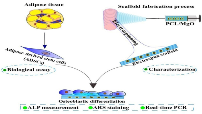
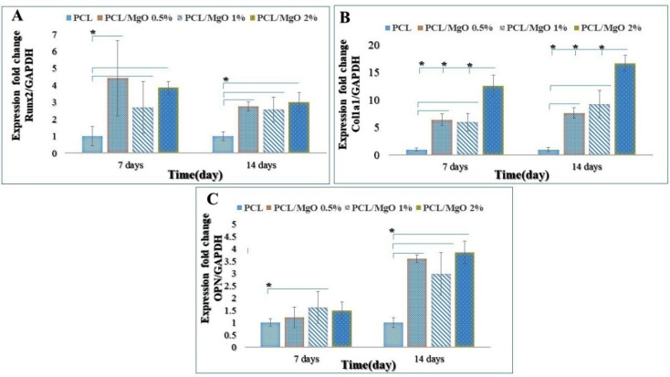
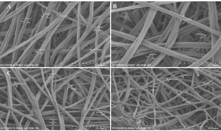
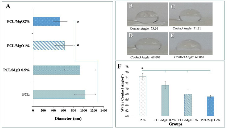
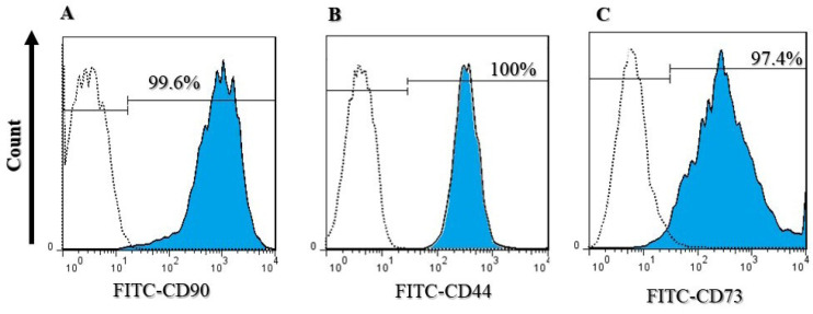
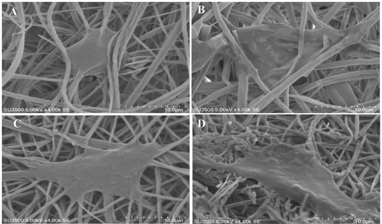
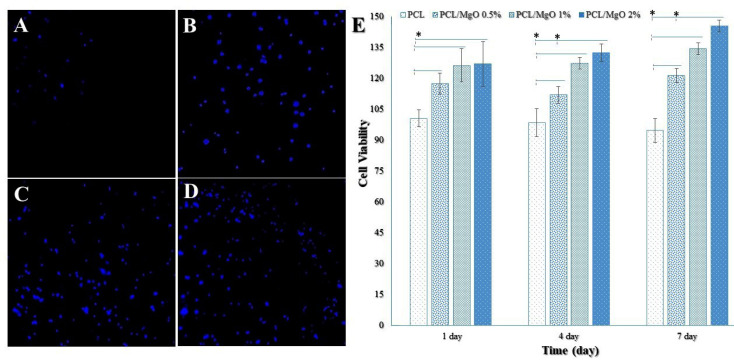
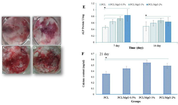
Recently, bone tissue engineering as a new strategy is used to repair and replace bone defects due to limitations in allograft and autograft methods. In this regard, we prepared nanofibrous scaffolds composed of polycaprolactone (PCL) and magnesium oxide (MgO) nanoparticles using the electrospinning technique for possible bone tissue engineering applications. The fabricated composites were characterized via scanning electron microscopy (SEM) imaging of scaffolds and seeded cells, water contact angle, DAPI staining, and MTT assay. Then osteogenic differentiation of adipose-derived mesenchymal stem cells cultured on this composite scaffold was determined by standard osteogenic marker tests, including alkaline phosphatase (ALP) activity, calcium deposition, and expression of osteogenic differentiation genes in the laboratory conditions. The SEM analysis demonstrated that the diameter of nanofibers significantly decreased from 1029.25±209.349 µm to 537.83+0.140 nm, with the increase of MgO concentration to 2% ( < 0.05). Initial adhesion and proliferation of the adipose-derived mesenchymal stem cells on MgO/PCL scaffolds were significantly enhanced with the increasing of MgO concentration ( < 0.05). The 2% MgO/PCL nanofibrous scaffold showed significant increase in ALP activity ( < 0.05) and osteogenic-related gene expressions (Col1a1 and OPN) ( < 0.05) in compared to pure PCL and (0, 0.5 and 1%) MgO/PCL scaffolds. According to the results, it was demonstrated that MgO/PCL composite nanofibers have considerable osteoinductive potential, and taking together adipose-derived mesenchymal stem cells-MgO/PCL composite nanofibers can be a proper bio-implant to usage for bone regenerative medicine applications. Future in vivo studies are needed to determine this composite therapeutic potential.

摘要

近年来,由于同种异体移植和自体移植方法存在局限性,骨组织工程作为一种新策略被用于修复和替代骨缺损。在这方面,我们采用静电纺丝技术制备了由聚己内酯(PCL)和氧化镁(MgO)纳米颗粒组成的纳米纤维支架,用于可能的骨组织工程应用。通过对支架和接种细胞的扫描电子显微镜(SEM)成像、水接触角、DAPI染色和MTT测定对制备的复合材料进行了表征。然后,通过标准的成骨标志物测试,包括碱性磷酸酶(ALP)活性、钙沉积以及在实验室条件下成骨分化基因的表达,来确定在这种复合支架上培养的脂肪来源间充质干细胞的成骨分化情况。SEM分析表明,随着MgO浓度增加到2%,纳米纤维直径从1029.25±209.349 µm显著减小至537.83+0.140 nm(P<0.05)。随着MgO浓度的增加,脂肪来源间充质干细胞在MgO/PCL支架上的初始黏附和增殖显著增强(P<0.05)。与纯PCL以及(0、0.5和1%)MgO/PCL支架相比,2% MgO/PCL纳米纤维支架的ALP活性(P<0.05)和成骨相关基因表达(Col1a1和OPN)(P<0.05)显著增加。根据结果表明,MgO/PCL复合纳米纤维具有相当大的骨诱导潜力,综合来看,脂肪来源间充质干细胞-MgO/PCL复合纳米纤维可以作为一种合适的生物植入物用于骨再生医学应用。未来需要进行体内研究以确定这种复合材料的治疗潜力。

https://cdn.ncbi.nlm.nih.gov/pmc/blobs/8d40/9012933/f871c79a6435/apb-12-142-g008.jpg
https://cdn.ncbi.nlm.nih.gov/pmc/blobs/8d40/9012933/945d60509557/apb-12-142-g001.jpg
https://cdn.ncbi.nlm.nih.gov/pmc/blobs/8d40/9012933/cb863c74d5d5/apb-12-142-g002.jpg
https://cdn.ncbi.nlm.nih.gov/pmc/blobs/8d40/9012933/1dc6ac309ba2/apb-12-142-g003.jpg
https://cdn.ncbi.nlm.nih.gov/pmc/blobs/8d40/9012933/73a8b905dc2d/apb-12-142-g004.jpg
https://cdn.ncbi.nlm.nih.gov/pmc/blobs/8d40/9012933/b43d77a769ea/apb-12-142-g005.jpg
https://cdn.ncbi.nlm.nih.gov/pmc/blobs/8d40/9012933/92b60b20120e/apb-12-142-g006.jpg
https://cdn.ncbi.nlm.nih.gov/pmc/blobs/8d40/9012933/e34fa04ff497/apb-12-142-g007.jpg
https://cdn.ncbi.nlm.nih.gov/pmc/blobs/8d40/9012933/f871c79a6435/apb-12-142-g008.jpg

相似文献

1
Osteogenic Differentiation Potential of Adipose-Derived Mesenchymal Stem Cells Cultured on Magnesium Oxide/Polycaprolactone Nanofibrous Scaffolds for Improving Bone Tissue Reconstruction.在氧化镁/聚己内酯纳米纤维支架上培养的脂肪来源间充质干细胞的成骨分化潜能用于改善骨组织重建
Adv Pharm Bull. 2022 Jan;12(1):142-154. doi: 10.34172/apb.2022.015. Epub 2020 Sep 22.
2
Morphological and Molecular Analysis of Osteoblasts Differentiated from Mesenchymal Stem Cells in Polycaprolactone/Magnesium Oxide/Graphene Oxide Scaffold.聚己内酯/氧化镁/氧化石墨烯支架中从间充质干细胞分化而来的成骨细胞的形态学和分子分析
Int J Organ Transplant Med. 2019;10(4):171-182.
3
PCL/Col I-based magnetic nanocomposite scaffold provides an osteoinductive environment for ADSCs in osteogenic cues-free media conditions.基于聚己内酯/ I型胶原的磁性纳米复合支架在无成骨信号的培养基条件下为脂肪来源干细胞提供了一个骨诱导环境。
Stem Cell Res Ther. 2022 Apr 4;13(1):143. doi: 10.1186/s13287-022-02816-0.
4
Nanofibrous Mineralized Electrospun Scaffold as a Substrate for Bone Tissue Regeneration.纳米纤维矿化电纺支架作为骨组织再生的基质
J Biomed Nanotechnol. 2016 Nov;12(11):2076-82. doi: 10.1166/jbn.2016.2306.
5
Adipose-derived stem cells-conditioned medium improved osteogenic differentiation of induced pluripotent stem cells when grown on polycaprolactone nanofibers.脂肪来源干细胞条件培养基在聚己内酯纳米纤维上培养时可提高诱导多能干细胞的成骨分化。
J Cell Physiol. 2019 Jul;234(7):10315-10323. doi: 10.1002/jcp.27697. Epub 2018 Oct 30.
6
Drug-eluting PCL/graphene oxide nanocomposite scaffolds for enhanced osteogenic differentiation of mesenchymal stem cells.载药聚己内酯/氧化石墨烯纳米复合支架促进间充质干细胞成骨分化。
Mater Sci Eng C Mater Biol Appl. 2020 Oct;115:111102. doi: 10.1016/j.msec.2020.111102. Epub 2020 May 20.
7
3D printed PLGA/MgO/PDA composite scaffold by low-temperature deposition manufacturing for bone tissue engineering applications.用于骨组织工程应用的低温沉积制造3D打印PLGA/MgO/PDA复合支架
Regen Ther. 2023 Nov 11;24:617-629. doi: 10.1016/j.reth.2023.09.015. eCollection 2023 Dec.
8
Synthesis of bone biocompatible implants using human adipose-derived mesenchymal stem cells (hADMSCs) and PCL/laminin scaffold substrate.利用人脂肪来源间充质干细胞(hADMSCs)和聚己内酯/层粘连蛋白支架基质合成骨生物相容性植入物。
Iran J Basic Med Sci. 2024;27(2):118-194. doi: 10.22038/IJBMS.2023.71307.15491.
9
In-situ polymerized polypyrrole nanoparticles immobilized poly(ε-caprolactone) electrospun conductive scaffolds for bone tissue engineering.原位聚合聚吡咯纳米粒子固定化聚(ε-己内酯)电纺导电支架用于骨组织工程
Mater Sci Eng C Mater Biol Appl. 2020 Sep;114:111056. doi: 10.1016/j.msec.2020.111056. Epub 2020 May 6.
10
Osteogenic differentiation potential of mesenchymal stem cells cultured on nanofibrous scaffold improved in the presence of pulsed electromagnetic field.在脉冲电磁场存在的情况下,培养在纳米纤维支架上的间充质干细胞的成骨分化潜能得到改善。
J Cell Physiol. 2018 Feb;233(2):1061-1070. doi: 10.1002/jcp.25962. Epub 2017 Jun 6.

引用本文的文献

1
Hydrogel-Based Scaffolds: Advancing Bone Regeneration Through Tissue Engineering.基于水凝胶的支架:通过组织工程促进骨再生
Gels. 2025 Feb 27;11(3):175. doi: 10.3390/gels11030175.
2
Advanced progress of adipose-derived stem cells-related biomaterials in maxillofacial regeneration.脂肪来源干细胞相关生物材料在颌面再生中的研究进展
Stem Cell Res Ther. 2025 Mar 5;16(1):110. doi: 10.1186/s13287-025-04191-y.
3
Advances in magnesium-containing bioceramics for bone repair.用于骨修复的含镁生物陶瓷的研究进展。

本文引用的文献

1
Morphological and Molecular Analysis of Osteoblasts Differentiated from Mesenchymal Stem Cells in Polycaprolactone/Magnesium Oxide/Graphene Oxide Scaffold.聚己内酯/氧化镁/氧化石墨烯支架中从间充质干细胞分化而来的成骨细胞的形态学和分子分析
Int J Organ Transplant Med. 2019;10(4):171-182.
2
Biological Response to Macroporous Chitosan-Agarose Bone Scaffolds Comprising Mg- and Zn-Doped Nano-Hydroxyapatite.生物对包含 Mg 和 Zn 掺杂纳米羟基磷灰石的大孔壳聚糖-琼脂糖骨支架的反应。
Int J Mol Sci. 2019 Aug 6;20(15):3835. doi: 10.3390/ijms20153835.
3
Betulin Promotes Differentiation of Human Osteoblasts In Vitro and Exerts an Osteoinductive Effect on the hFOB 1.19 Cell Line Through Activation of JNK, ERK1/2, and mTOR Kinases.
Biomater Transl. 2024 Mar 28;5(1):3-20. doi: 10.12336/biomatertransl.2024.01.002. eCollection 2024.
4
Enhanced osteogenesis and bactericidal performance with additively manufactured MgO and Cu-added CpTi for load-bearing implants.通过增材制造的MgO和添加Cu的纯钛实现增强的成骨和杀菌性能用于承重植入物。
Int J Bioprint. 2023 Oct 15;9(6). doi: 10.36922/ijb.1167. Epub 2023 Oct 11.
5
The beneficial effects of simultaneous supplementation of Lactobacillus reuteri and calcium fluoride nanoparticles on ovariectomy-induced osteoporosis.同时补充罗伊氏乳杆菌和氟化钙纳米粒子对去卵巢诱导骨质疏松症的有益影响。
BMC Complement Med Ther. 2023 Sep 26;23(1):340. doi: 10.1186/s12906-023-04167-6.
6
How Mechanical and Physicochemical Material Characteristics Influence Adipose-Derived Stem Cell Fate.机械和物理化学材料特性如何影响脂肪来源干细胞命运。
Int J Mol Sci. 2023 Feb 10;24(4):3551. doi: 10.3390/ijms24043551.
7
Electrospun Polycaprolactone Nanofibers: Current Research and Applications in Biomedical Application.静电纺聚己内酯纳米纤维:生物医学应用中的当前研究与应用
Adv Pharm Bull. 2022 Aug;12(4):658-672. doi: 10.34172/apb.2022.070. Epub 2021 Oct 3.
8
Improved osteogenic differentiation by extremely low electromagnetic field exposure: possible application for bone engineering.极低频电磁场暴露促进成骨分化:在骨工程中的可能应用。
Histochem Cell Biol. 2022 Oct;158(4):369-381. doi: 10.1007/s00418-022-02126-9. Epub 2022 Jun 25.
9
Strontium doped bioglass incorporated hydrogel-based scaffold for amplified bone tissue regeneration.锶掺杂生物玻璃复合水凝胶基支架用于增强骨组织再生。
Sci Rep. 2022 Jun 17;12(1):10160. doi: 10.1038/s41598-022-14329-0.
10
Inorganic Nanoparticles in Bone Healing Applications.骨愈合应用中的无机纳米颗粒
Pharmaceutics. 2022 Mar 31;14(4):770. doi: 10.3390/pharmaceutics14040770.
桦木醇促进体外人成骨细胞分化,并通过激活 JNK、ERK1/2 和 mTOR 激酶对 hFOB 1.19 细胞系发挥成骨诱导作用。
Molecules. 2019 Jul 19;24(14):2637. doi: 10.3390/molecules24142637.
4
In vitro induction of odontogenic activity of human dental pulp stem cells by white Portland cement enriched with zirconium oxide and zinc oxide components.富含氧化锆和氧化锌成分的白色波特兰水泥对人牙髓干细胞牙源性活性的体外诱导作用
J Dent Res Dent Clin Dent Prospects. 2019 Winter;13(1):3-10. doi: 10.15171/joddd.2019.001. Epub 2019 Apr 24.
5
Biological behavior of the curcumin incorporated chitosan/poly(vinyl alcohol) nanofibers for biomedical applications.载姜黄素壳聚糖/聚乙烯醇纳米纤维的生物行为及其在生物医学中的应用。
J Cell Biochem. 2019 Sep;120(9):15410-15421. doi: 10.1002/jcb.28808. Epub 2019 May 8.
6
Regulation of Proliferation, Differentiation and Functions of Osteoblasts by Runx2.Runx2 对成骨细胞增殖、分化和功能的调节。
Int J Mol Sci. 2019 Apr 4;20(7):1694. doi: 10.3390/ijms20071694.
7
Comparison of osteogenic differentiation capacity in mesenchymal stem cells derived from human amniotic membrane (AM), umbilical cord (UC), chorionic membrane (CM), and decidua (DC).人羊膜(AM)、脐带(UC)、绒毛膜(CM)和蜕膜(DC)来源的间充质干细胞成骨分化能力的比较。
Cell Biosci. 2019 Feb 11;9:17. doi: 10.1186/s13578-019-0281-3. eCollection 2019.
8
In vitro osteogenic differentiation potential of the human induced pluripotent stem cells augments when grown on Graphene oxide-modified nanofibers.人诱导多能干细胞在氧化石墨烯修饰的纳米纤维上生长时,其体外成骨分化潜能增强。
Gene. 2019 May 15;696:72-79. doi: 10.1016/j.gene.2019.02.028. Epub 2019 Feb 15.
9
Cellular Response to Surface Morphology: Electrospinning and Computational Modeling.细胞对表面形态的反应:静电纺丝与计算建模
Front Bioeng Biotechnol. 2018 Oct 24;6:155. doi: 10.3389/fbioe.2018.00155. eCollection 2018.
10
The Clinical Trials of Mesenchymal Stem Cell Therapy in Skin Diseases: An Update and Concise Review.间充质干细胞疗法在皮肤病中的临床试验:最新进展与简要综述
Curr Stem Cell Res Ther. 2019;14(1):22-33. doi: 10.2174/1574888X13666180913123424.