Hu Likun, Zhang Ting, Liu Dong, Guan Guiwen, Huang Jian, Proksch Peter, Chen Xiangmei, Lin Wenhan
State Key Laboratory of Natural and Biomimetic Drugs, Peking University Beijing 100191 P. R. China
Department of Microbiology and Infectious Disease Center, School of Basic Medical Sciences, Peking University Health Science Center Beijing 100191 P. R. China
RSC Adv. 2019 Jun 25;9(34):19855-19868. doi: 10.1039/c9ra03640g. eCollection 2019 Jun 19.
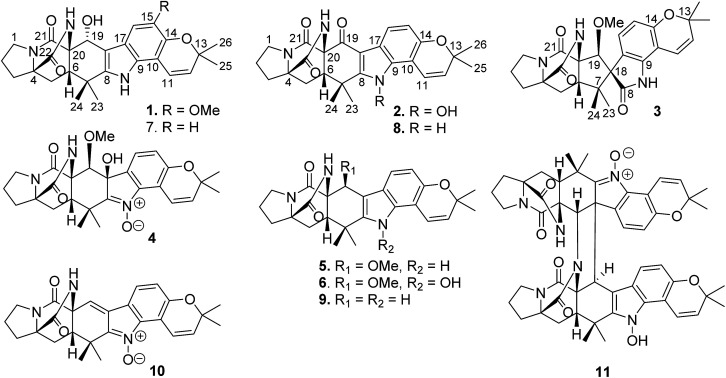
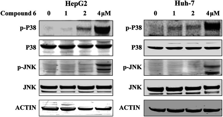
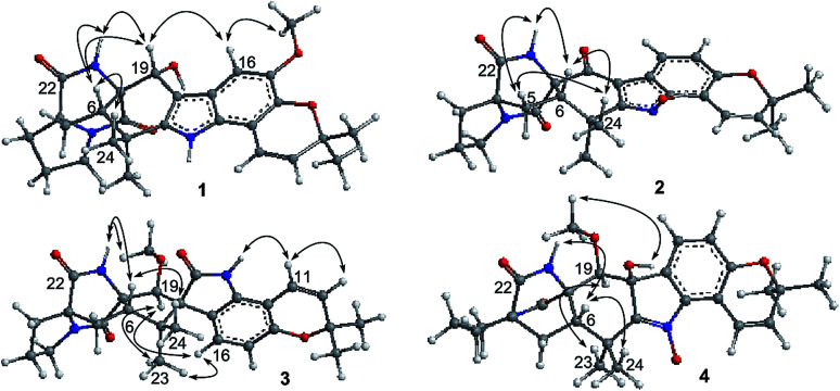
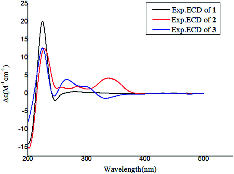
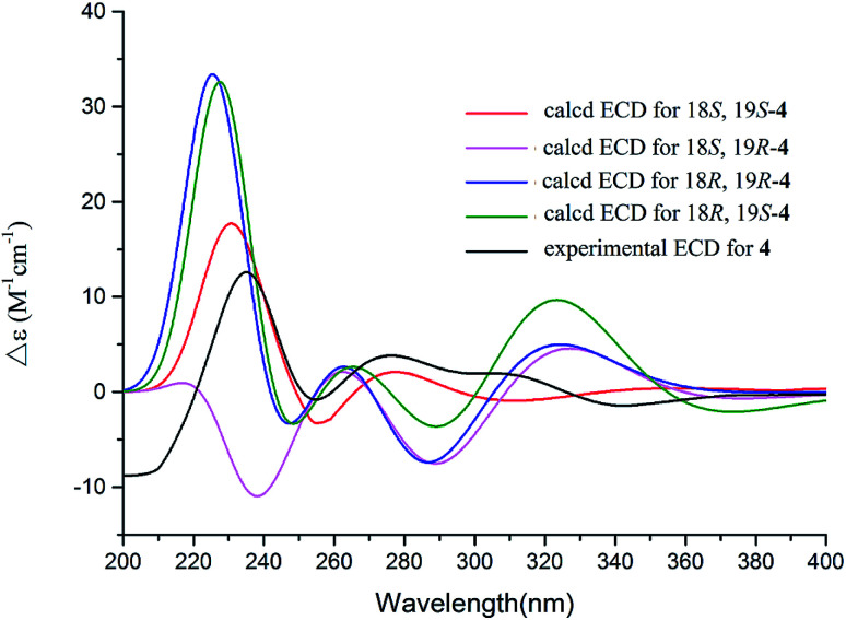
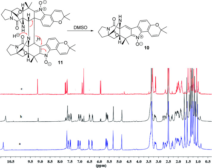
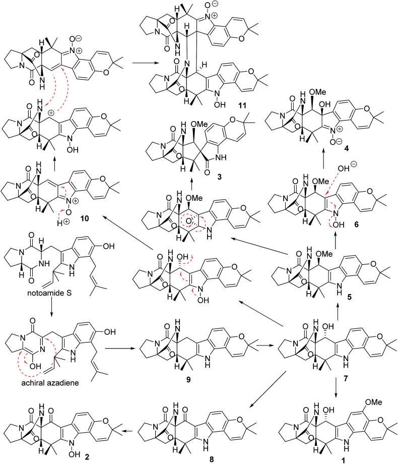
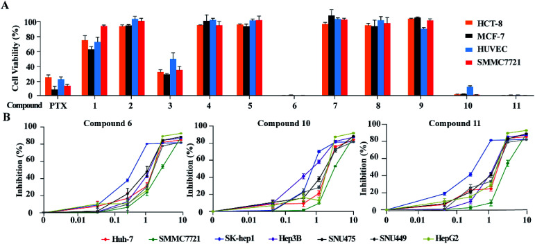
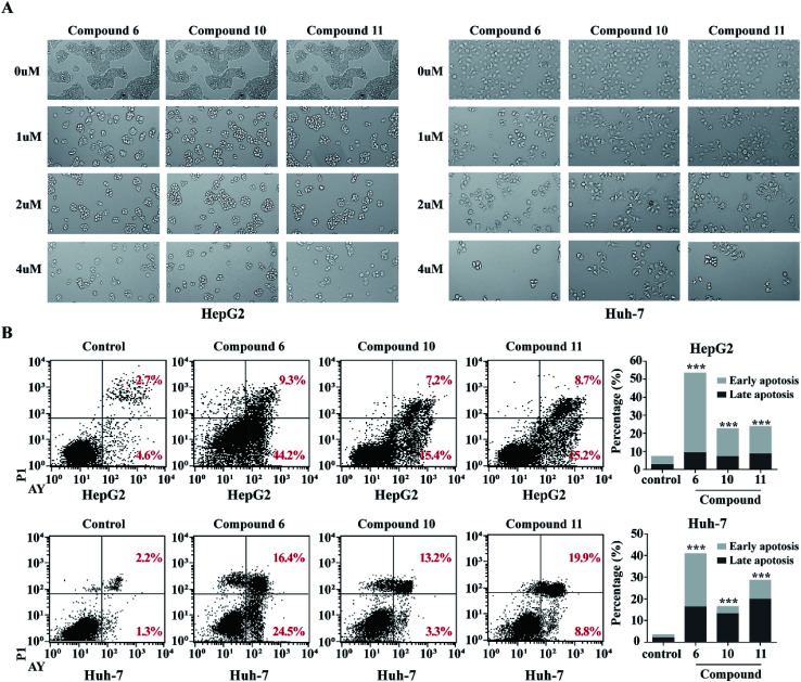
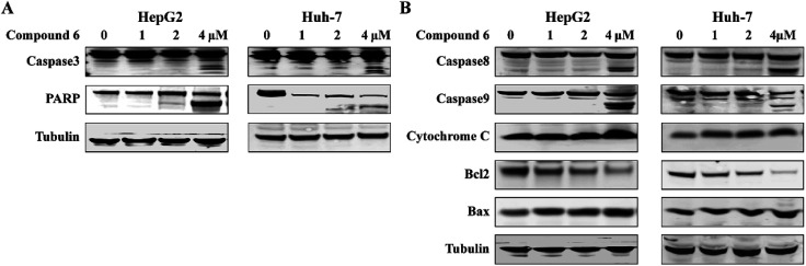
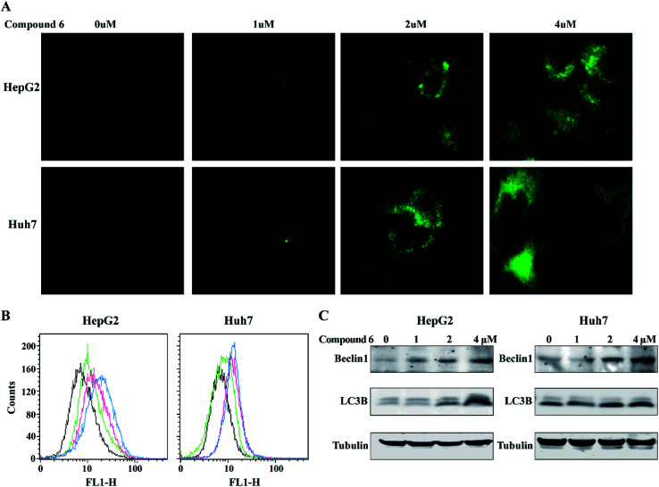
Bioassay-guided fractionation of a coral-associated fungus LZDX-32-15 resulted in the isolation of eleven notoamide-type alkaloids, including four new congeners, namely notoamides W-Z (1-4). The structures of the new alkaloids were determined by extensive analyses of spectroscopic data (1D and 2D NMR, HRESIMS), while ECD data were used for the configurational assignment. Three alkaloids (6, 10, 11) exerted potent inhibition against a panel of hepatocellular carcinoma (HCC) cell lines with IC values ranging from 0.42 to 3.39 μM, that are comparable to the data for paclitaxel. Notoamide G (6) inhibited the viability of HepG2 and Huh-7 cells both apoptosis and autophagy pathways. Notoamide G activated the expression of caspase-3, caspase-8, and caspase-9, in association with the degradation of the downstream gene PARP in a dose-dependent manner, suggesting that notoamide G induced apoptosis a mitochondrial pathway and a dead receptor-mediated pathway. In addition, notoamide G increased the autophagic vacuole in both HepG2 and Huh-7 cells in a dose-dependent manner after 24 h through the significant upregulation of the key proteins Beclin1 and LC3B. Further investigation revealed that notoamide G promoted P38 and JNK phosphorylation, whereas the total protein of P-38 and JNK was slightly influenced. Accordingly, the antitumor proliferation of notoamide G in HCC cells was mechanistically mediated by apoptosis and autophagy through a P38/JNK signaling pathway, while notoamide G was considered as a potent lead for further development as an antitumor agent.
对一种与珊瑚相关的真菌LZDX - 32 - 15进行生物测定导向的分级分离,得到了11种诺托酰胺型生物碱,包括4个新的同系物,即诺托酰胺W - Z(1 - 4)。通过对光谱数据(一维和二维核磁共振、高分辨电喷雾电离质谱)的广泛分析确定了新生物碱的结构,同时利用电子圆二色光谱数据进行构型归属。三种生物碱(6、10、11)对一组肝癌(HCC)细胞系具有强效抑制作用,IC值范围为0.42至3.39 μM,与紫杉醇的数据相当。诺托酰胺G(6)抑制HepG2和Huh - 7细胞的活力,涉及凋亡和自噬途径。诺托酰胺G以剂量依赖的方式激活caspase - 3、caspase - 8和caspase - 9的表达,并伴有下游基因PARP的降解,表明诺托酰胺G通过线粒体途径和死亡受体介导的途径诱导凋亡。此外,诺托酰胺G在24小时后通过显著上调关键蛋白Beclin1和LC3B,以剂量依赖的方式增加了HepG2和Huh - 7细胞中的自噬泡。进一步研究表明,诺托酰胺G促进P38和JNK磷酸化,而P - 38和JNK的总蛋白受到轻微影响。因此,诺托酰胺G在肝癌细胞中的抗肿瘤增殖作用是通过P38/JNK信号通路由凋亡和自噬机制介导的,而诺托酰胺G被认为是一种有潜力的进一步开发为抗肿瘤药物的先导化合物。