• 文献检索
  • 文档翻译
  • 深度研究
  • 学术资讯
  • Suppr Zotero 插件Zotero 插件
  • 邀请有礼
  • 套餐&价格
  • 历史记录
应用&插件
Suppr Zotero 插件Zotero 插件浏览器插件Mac 客户端Windows 客户端微信小程序
定价
高级版会员购买积分包购买API积分包
服务
文献检索文档翻译深度研究API 文档MCP 服务
关于我们
关于 Suppr公司介绍联系我们用户协议隐私条款
关注我们

Suppr 超能文献

核心技术专利:CN118964589B侵权必究
粤ICP备2023148730 号-1Suppr @ 2026

文献检索

告别复杂PubMed语法,用中文像聊天一样搜索,搜遍4000万医学文献。AI智能推荐,让科研检索更轻松。

立即免费搜索

文件翻译

保留排版,准确专业,支持PDF/Word/PPT等文件格式,支持 12+语言互译。

免费翻译文档

深度研究

AI帮你快速写综述,25分钟生成高质量综述,智能提取关键信息,辅助科研写作。

立即免费体验

微波等离子体辅助硅纳米颗粒:对癌细胞的细胞毒性、分子及数值反应

Microwave plasma-assisted silicon nanoparticles: cytotoxic, molecular, and numerical responses against cancer cells.

作者信息

Wahab Rizwan, Khan Farheen, Gupta Anoop, Wiggers Hartmut, Saquib Quaiser, Faisal Mohammad, Ansari Sabiha Mahmood

机构信息

Zoology Department, College of Science, King Saud University P. O. Box 2455 Riyadh 11451 Saudi Arabia

Chemistry Department, Faculty of Science, Taibah University Yanbu Saudi Arabia

出版信息

RSC Adv. 2019 Apr 30;9(23):13336-13347. doi: 10.1039/c8ra10185j. eCollection 2019 Apr 25.

DOI:10.1039/c8ra10185j
PMID:35520784
原文链接:https://pmc.ncbi.nlm.nih.gov/articles/PMC9063978/
Abstract

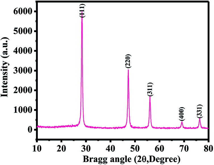
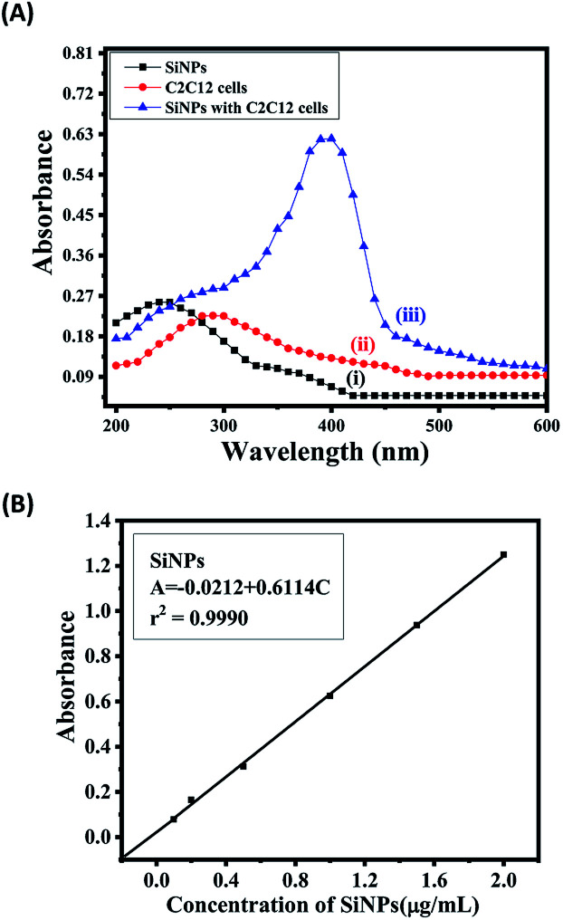
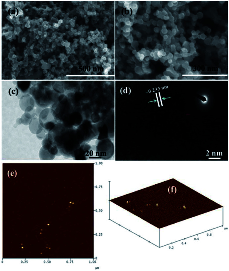
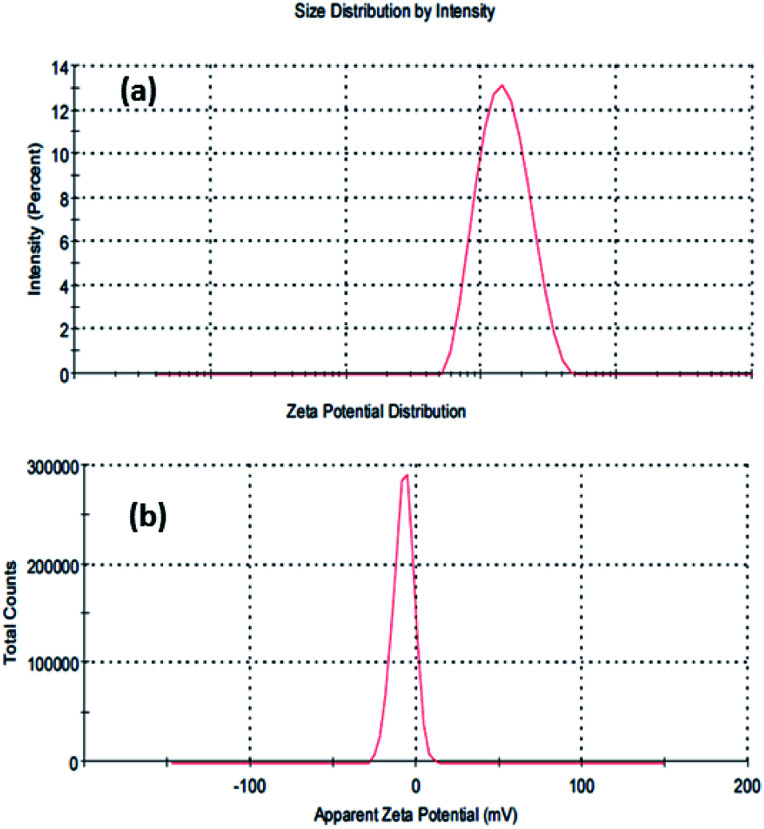
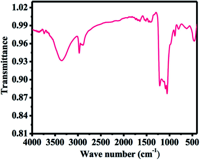
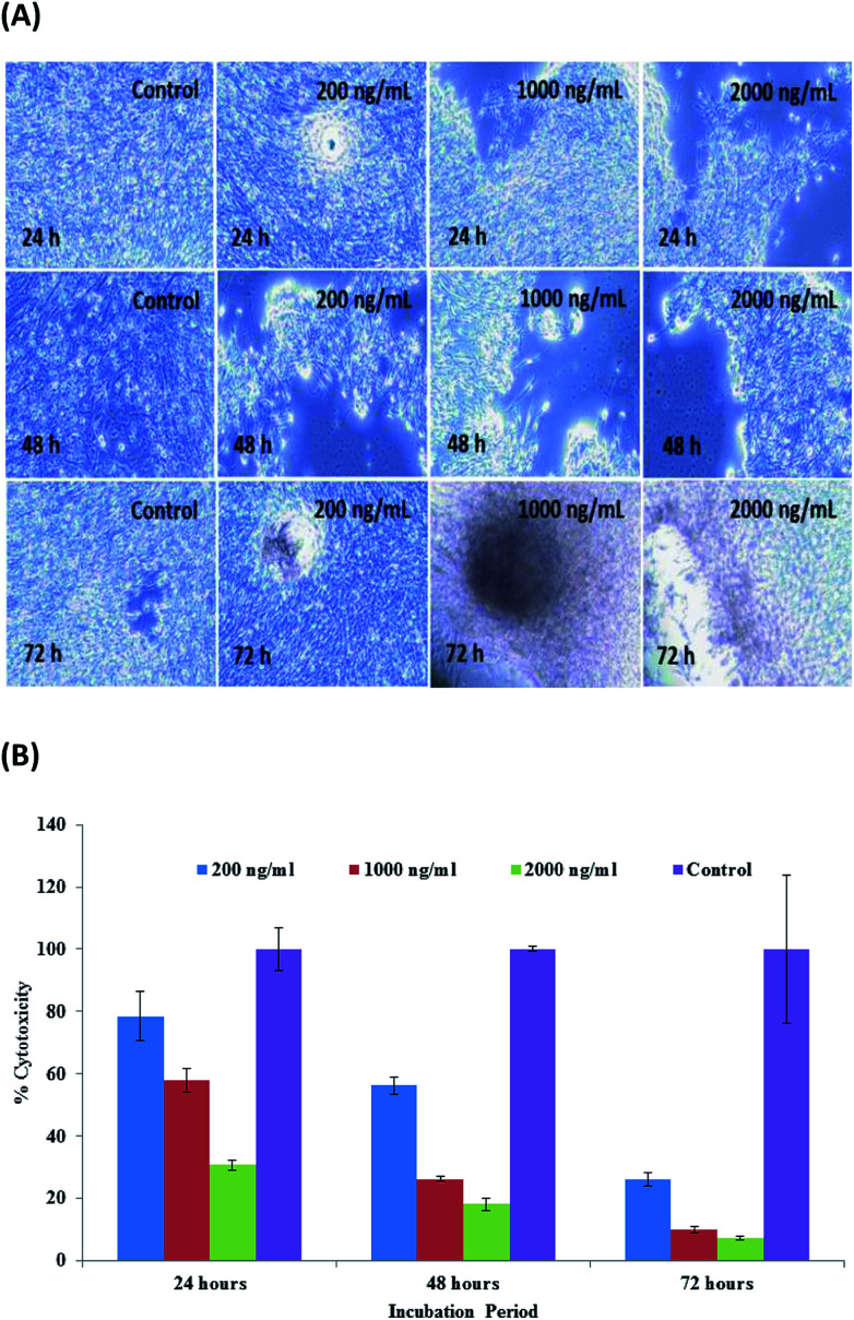
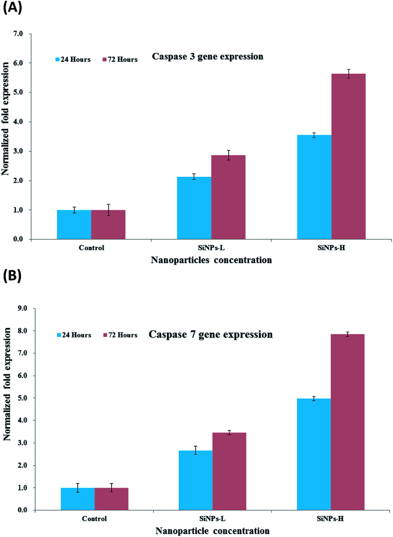
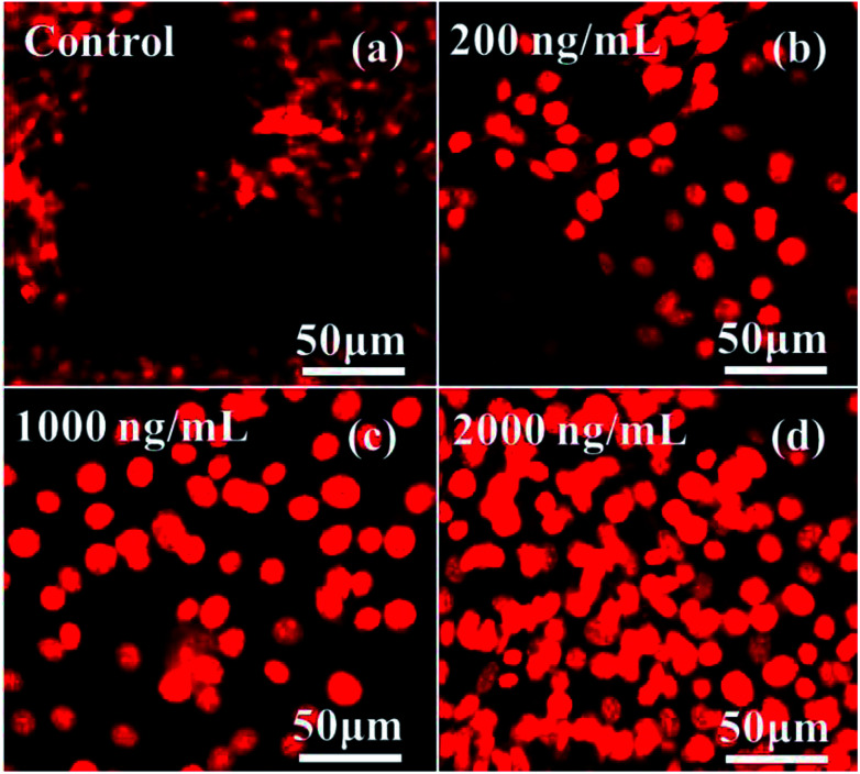
Silicon nanoparticles (SiNPs), which have a special place in material science due to their strong luminescent property and wide applicability in various physicochemical arenas, such as solar cells and LEDs, were synthesised by a microwave plasma-assisted process using an argon-silane mixture. Several characterization tools were applied to check the crystallinity (XRD) and morphological (FESEM, TEM, ∼20 ± 2 nm size) and topographical (AFM, ∼20 nm) details of the NPs. The high-purity SiNPs were applied on myoblast cancer cells to investigate the reactivity of the NPs at different doses (200, 1000 and 2000 ng mL) for different incubation periods (24 h, 48 h & 72 h). The MTT assay was utilized to determine the percentage of viable and non-viable cells, while the cell organization was observed microscopy and CLSM. Additionally, the molecular responses (RT-PCR), such as apoptosis, were analyzed in presence of caspase 3 and 7, and the results showed an upregulation with SiNPs. To validate the obtained data, analytical studies were also performed for the SiNPs statistical analysis and the most reliable data values were evaluated and acceptable as per the ICH guidelines.

摘要

硅纳米颗粒(SiNPs)因其强发光特性以及在太阳能电池和发光二极管等各种物理化学领域的广泛适用性,在材料科学中占有特殊地位。通过使用氩 - 硅烷混合物的微波等离子体辅助工艺合成了硅纳米颗粒。应用了多种表征工具来检查纳米颗粒的结晶度(XRD)、形态(FESEM、TEM,尺寸约为20±2纳米)和形貌(AFM,约20纳米)细节。将高纯度的硅纳米颗粒应用于成肌细胞瘤细胞,以研究不同剂量(200、1000和2000纳克/毫升)的纳米颗粒在不同孵育时间(24小时、48小时和72小时)的反应性。利用MTT法测定存活和非存活细胞的百分比,同时通过显微镜和共聚焦激光扫描显微镜观察细胞组织。此外,在存在半胱天冬酶3和7的情况下分析了诸如凋亡等分子反应(RT-PCR),结果显示硅纳米颗粒使其上调。为了验证所获得的数据,还对硅纳米颗粒进行了分析研究,进行了统计分析,并根据国际人用药品注册技术协调会(ICH)指南评估了最可靠的数据值并认为其可接受。

https://cdn.ncbi.nlm.nih.gov/pmc/blobs/83e0/9063978/d81f02cc2908/c8ra10185j-f8.jpg
https://cdn.ncbi.nlm.nih.gov/pmc/blobs/83e0/9063978/bb1b090eee51/c8ra10185j-f1.jpg
https://cdn.ncbi.nlm.nih.gov/pmc/blobs/83e0/9063978/03456dacb1c6/c8ra10185j-f2.jpg
https://cdn.ncbi.nlm.nih.gov/pmc/blobs/83e0/9063978/3fd53d6964e4/c8ra10185j-f3.jpg
https://cdn.ncbi.nlm.nih.gov/pmc/blobs/83e0/9063978/b6db9610f735/c8ra10185j-f4.jpg
https://cdn.ncbi.nlm.nih.gov/pmc/blobs/83e0/9063978/4fb68c710fd0/c8ra10185j-f5.jpg
https://cdn.ncbi.nlm.nih.gov/pmc/blobs/83e0/9063978/7d0d2e215799/c8ra10185j-f6.jpg
https://cdn.ncbi.nlm.nih.gov/pmc/blobs/83e0/9063978/9cb53508f35c/c8ra10185j-f7.jpg
https://cdn.ncbi.nlm.nih.gov/pmc/blobs/83e0/9063978/d81f02cc2908/c8ra10185j-f8.jpg

相似文献

1
Microwave plasma-assisted silicon nanoparticles: cytotoxic, molecular, and numerical responses against cancer cells.微波等离子体辅助硅纳米颗粒:对癌细胞的细胞毒性、分子及数值反应
RSC Adv. 2019 Apr 30;9(23):13336-13347. doi: 10.1039/c8ra10185j. eCollection 2019 Apr 25.
2
Hematite iron oxide nanoparticles: apoptosis of myoblast cancer cells and their arithmetical assessment.赤铁矿氧化铁纳米颗粒:成肌细胞瘤细胞的凋亡及其算术评估
RSC Adv. 2018 Jul 9;8(44):24750-24759. doi: 10.1039/c8ra02613k.
3
Statistical analysis of gold nanoparticle-induced oxidative stress and apoptosis in myoblast (C2C12) cells.金纳米颗粒诱导成肌细胞(C2C12)氧化应激和凋亡的统计分析
Colloids Surf B Biointerfaces. 2014 Nov 1;123:664-72. doi: 10.1016/j.colsurfb.2014.10.012. Epub 2014 Oct 12.
4
Synthesis of D-mannose capped silicon nanoparticles and their interactions with MCF-7 human breast cancerous cells.D-甘露糖封端硅纳米粒子的合成及其与 MCF-7 人乳腺癌细胞的相互作用。
ACS Appl Mater Interfaces. 2013 Aug 14;5(15):7384-91. doi: 10.1021/am4017126. Epub 2013 Jul 16.
5
Preparation and characterization of decyl-terminated silicon nanoparticles encapsulated in lipid nanocapsules.癸基封端硅纳米粒子的制备及脂质纳米胶囊包封的表征。
Langmuir. 2013 Oct 15;29(41):12688-96. doi: 10.1021/la4029468. Epub 2013 Oct 1.
6
One-Pot Microwave Synthesis of Water-Dispersible, High Fluorescence Silicon Nanoparticles and Their Imaging Applications in Vitro and in Vivo.一锅微波法合成水分散性高荧光硅纳米颗粒及其在体内外成像应用。
Anal Chem. 2016 Dec 6;88(23):11631-11638. doi: 10.1021/acs.analchem.6b03209. Epub 2016 Nov 14.
7
In vitro cellular behaviors and toxicity assays of small-sized fluorescent silicon nanoparticles.小尺寸荧光硅纳米颗粒的体外细胞行为和毒性检测。
Nanoscale. 2017 Jun 8;9(22):7602-7611. doi: 10.1039/c7nr00530j.
8
Two-photon-excited tumor cell fluorescence targeted imaging based on transferrin-functionalized silicon nanoparticles.基于转铁蛋白功能化硅纳米粒子的双光子激发肿瘤细胞荧光靶向成像。
Spectrochim Acta A Mol Biomol Spectrosc. 2022 Feb 15;267(Pt 1):120450. doi: 10.1016/j.saa.2021.120450. Epub 2021 Oct 6.
9
Two distinct cellular pathways leading to endothelial cell cytotoxicity by silica nanoparticle size.两种不同的细胞通路导致二氧化硅纳米颗粒大小引起的内皮细胞细胞毒性。
J Nanobiotechnology. 2019 Feb 5;17(1):24. doi: 10.1186/s12951-019-0456-4.
10
Silica nanoparticles alleviate cadmium toxicity in rice cells: Mechanisms and size effects.二氧化硅纳米颗粒减轻水稻细胞中的镉毒性:作用机制及尺寸效应
Environ Pollut. 2017 Sep;228:363-369. doi: 10.1016/j.envpol.2017.05.014. Epub 2017 May 25.

引用本文的文献

1
The toxicity of SiO NPs on cell proliferation and cellular uptake of human lung fibroblastic cell line during the variation of calcination temperature and its modeling by artificial neural network.在煅烧温度变化期间,二氧化硅纳米颗粒对人肺成纤维细胞系细胞增殖和细胞摄取的毒性及其人工神经网络建模
J Environ Health Sci Eng. 2021 Apr 30;19(1):985-995. doi: 10.1007/s40201-021-00663-4. eCollection 2021 Jun.
2
Zinc oxide nanoparticles using plant Lawsonia inermis and their mosquitocidal, antimicrobial, anticancer applications showing moderate side effects.利用植物指甲花制备的氧化锌纳米粒子及其具有中度副作用的杀蚊、抗菌、抗癌应用。
Sci Rep. 2021 Apr 23;11(1):8837. doi: 10.1038/s41598-021-88164-0.

本文引用的文献

1
Resonant silicon nanoparticles for enhancement of light absorption and photoluminescence from hybrid perovskite films and metasurfaces.用于增强混合钙钛矿薄膜和超表面光吸收和光致发光的共振硅纳米颗粒。
Nanoscale. 2017 Aug 31;9(34):12486-12493. doi: 10.1039/c7nr01631j.
2
Functional Films from Silica/Polymer Nanoparticles.二氧化硅/聚合物纳米颗粒制成的功能薄膜
Materials (Basel). 2014 May 15;7(5):3881-3900. doi: 10.3390/ma7053881.
3
Mesoporous Silica Nanoparticles as Drug Delivery Vehicles in Cancer.介孔二氧化硅纳米颗粒作为癌症治疗中的药物递送载体
Nanomaterials (Basel). 2017 Jul 22;7(7):189. doi: 10.3390/nano7070189.
4
One-Pot Synthesis of Fluorescent Silicon Nanoparticles for Sensitive and Selective Determination of 2,4,6-Trinitrophenol in Aqueous Solution.一锅法合成荧光硅纳米粒子用于水溶液中 2,4,6-三硝基苯酚的灵敏选择性测定。
Anal Chem. 2017 Mar 7;89(5):3001-3008. doi: 10.1021/acs.analchem.6b04509. Epub 2017 Feb 21.
5
Cytotoxicity control of silicon nanoparticles by biopolymer coating and ultrasound irradiation for cancer theranostic applications.通过生物聚合物涂层和超声辐射控制硅纳米颗粒的细胞毒性,用于癌症治疗应用。
Nanotechnology. 2017 Mar 10;28(10):105102. doi: 10.1088/1361-6528/aa5b7c.
6
Cytotoxicity and DNA damage in mouse macrophages exposed to silica nanoparticles.暴露于二氧化硅纳米颗粒的小鼠巨噬细胞中的细胞毒性和DNA损伤。
Genet Mol Res. 2016 Aug 30;15(3):gmr9005. doi: 10.4238/gmr.15039005.
7
Biomimetic Preparation and Dual-Color Bioimaging of Fluorescent Silicon Nanoparticles.仿生制备及荧光硅纳米粒子的双色生物成像。
J Am Chem Soc. 2015 Nov 25;137(46):14726-32. doi: 10.1021/jacs.5b08685. Epub 2015 Nov 12.
8
Cytotoxicity of various types of gold-mesoporous silica nanoparticles in human breast cancer cells.不同类型的金-介孔二氧化硅纳米颗粒对人乳腺癌细胞的细胞毒性
Int J Nanomedicine. 2015 Sep 30;10:6075-87. doi: 10.2147/IJN.S90887. eCollection 2015.
9
Evaluation of silica nanoparticle binding to major human blood proteins.二氧化硅纳米颗粒与主要人体血液蛋白结合情况的评估。
Nanoscale Res Lett. 2014 Dec;9(1):2493. doi: 10.1186/1556-276X-9-668. Epub 2014 Dec 11.
10
Toxicity of silica nanoparticles depends on size, dose, and cell type.二氧化硅纳米颗粒的毒性取决于其大小、剂量和细胞类型。
Nanomedicine. 2015 Aug;11(6):1407-16. doi: 10.1016/j.nano.2015.03.004. Epub 2015 Mar 25.