Ocular and Stem Cell Translational Research Section, National Eye Institute, NIH, Bethesda, MD 20892.
Information Resources Technology Branch, National Center for Advancing Translational Sciences, NIH, Bethesda, MD 20892.
Proc Natl Acad Sci U S A. 2022 May 10;119(19):e2117553119. doi: 10.1073/pnas.2117553119. Epub 2022 May 6.
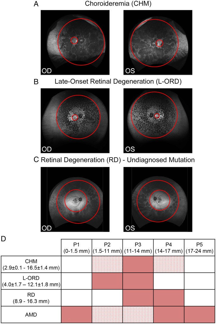
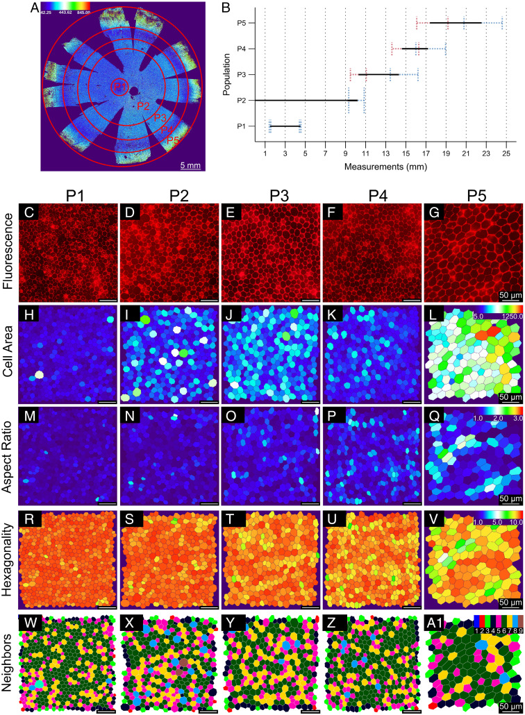
Regional phenotypic and functional differences in the retinal pigment epithelium (RPE) monolayer have been suggested to account for regional susceptibility in ocular diseases such as age-related macular degeneration (AMD), late-onset retinal degeneration (L-ORD), and choroideremia (CHM). However, a comprehensive description of human topographical RPE diversity is not yet available, thus limiting the understanding of regional RPE diversity and degenerative disease sensitivity in the eye. To develop a complete morphometric RPE map of the human eye, artificial intelligence–based software was trained to recognize, segment, and analyze RPE borders. Five statistically different, concentric RPE subpopulations (P1 to P5) were identified using cell area as a parameter, including a subpopulation (P4) with cell area comparable to that of macular cells in the far periphery of the eye. This work provides a complete reference map of human RPE subpopulations and their location in the eye. In addition, the analysis of cadaver non-AMD and AMD eyes and ultra-widefield fundus images of patients revealed differential vulnerability of the five RPE subpopulations to different retinal diseases.
已有研究表明,视网膜色素上皮(RPE)单层的区域性表型和功能差异可解释眼部疾病(如年龄相关性黄斑变性(AMD)、晚发性视网膜变性(L-ORD)和脉络膜视网膜变性(CHM))的区域性易感性。然而,目前尚缺乏对人类 RPE 拓扑多样性的全面描述,这限制了我们对眼部区域 RPE 多样性和退行性疾病敏感性的理解。为了开发完整的人类眼部 RPE 形态计量图谱,我们利用人工智能软件对 RPE 边界进行识别、分割和分析。本研究使用细胞面积作为参数,发现了 5 个具有统计学差异的同心 RPE 亚群(P1 到 P5),其中一个亚群(P4)的细胞面积与眼远周边部黄斑细胞相当。本工作提供了人类 RPE 亚群及其在眼部位置的完整参考图谱。此外,对非 AMD 尸检眼和 AMD 眼以及患者的超广角眼底图像进行分析,发现这 5 个 RPE 亚群对不同的视网膜疾病具有不同的易感性。