Brown Emma L, Lefebvre Thierry L, Sweeney Paul W, Stolz Bernadette J, Gröhl Janek, Hacker Lina, Huang Ziqiang, Couturier Dominique-Laurent, Harrington Heather A, Byrne Helen M, Bohndiek Sarah E
Department of Physics, University of Cambridge, JJ Thomson Avenue, Cambridge CB3 0HE, UK.
Cancer Research UK Cambridge Institute, University of Cambridge, Robinson Way, Cambridge CB2 0RE, UK.
Photoacoustics. 2022 Apr 20;26:100357. doi: 10.1016/j.pacs.2022.100357. eCollection 2022 Jun.
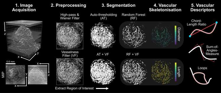
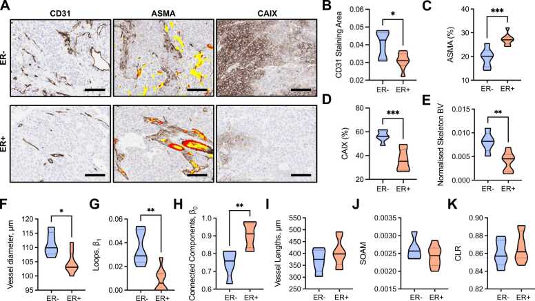
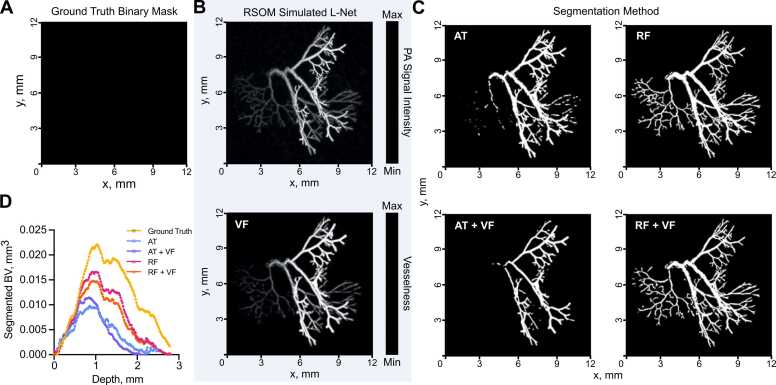
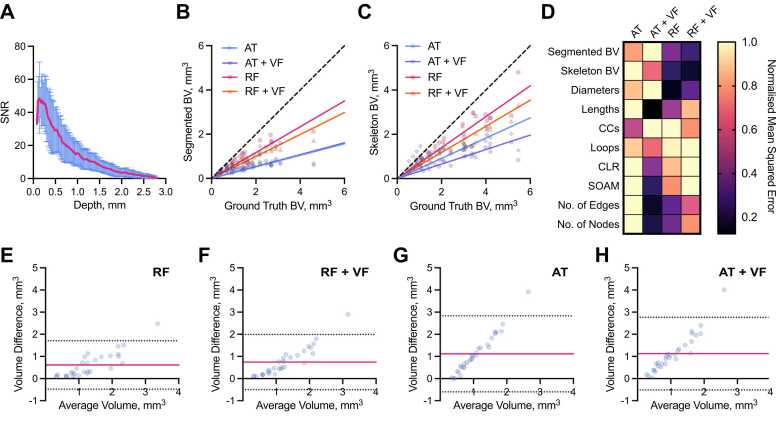
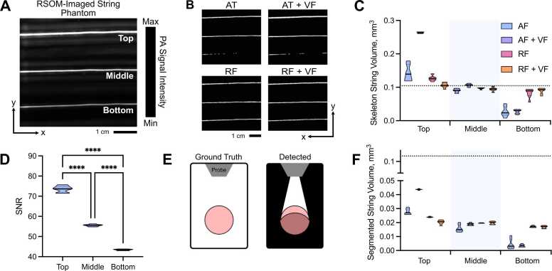
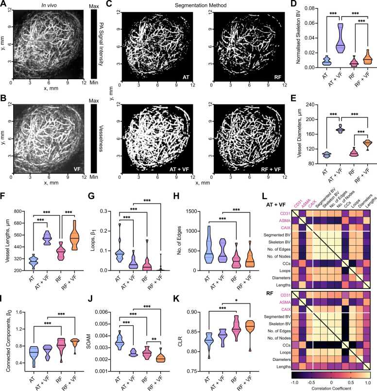
Mesoscopic photoacoustic imaging (PAI) enables non-invasive visualisation of tumour vasculature. The visual or semi-quantitative 2D measurements typically applied to mesoscopic PAI data fail to capture the 3D vessel network complexity and lack robust ground truths for assessment of accuracy. Here, we developed a pipeline for quantifying 3D vascular networks captured using mesoscopic PAI and tested the preservation of blood volume and network structure with topological data analysis. Ground truth data of synthetic vasculatures and a string phantom indicated that learning-based segmentation best preserves vessel diameter and blood volume at depth, while rule-based segmentation with vesselness image filtering accurately preserved network structure in superficial vessels. Segmentation of vessels in breast cancer patient-derived xenografts (PDXs) compared favourably to immunohistochemistry. Furthermore, our findings underscore the importance of validating segmentation methods when applying mesoscopic PAI as a tool to evaluate vascular networks .
介观光声成像(PAI)能够对肿瘤血管系统进行无创可视化。通常应用于介观PAI数据的视觉或半定量二维测量方法无法捕捉三维血管网络的复杂性,并且缺乏用于评估准确性的可靠基准真值。在此,我们开发了一种用于量化使用介观PAI捕获的三维血管网络的流程,并通过拓扑数据分析测试了血容量和网络结构的保存情况。合成血管和线状体模的基准真值数据表明,基于学习的分割方法最能在深度上保留血管直径和血容量,而使用血管增强图像滤波的基于规则的分割方法能准确保留浅表血管中的网络结构。对乳腺癌患者来源的异种移植瘤(PDX)中的血管进行分割,其效果优于免疫组织化学。此外,我们的研究结果强调了在将介观PAI用作评估血管网络的工具时验证分割方法的重要性。