Shenzhen Eye Institute, Shenzhen Eye Hospital Affiliated to Jinan University, Shenzhen, Guangdong, China.
School of Ophthalmology, Optometry, Shenzhen Eye Hospital, Shenzhen University, Shenzhen, China.
Transl Vis Sci Technol. 2022 May 2;11(5):12. doi: 10.1167/tvst.11.5.12.
The purpose of this study was to explore the therapeutic effect of human umbilical cord mesenchymal stem cell (HUMSC) transplantation alone or assisted with ultrasound targeted microbubble destruction (UTMD) on optic neuropathy in a novel and practical model of experimental glaucoma in rabbits.
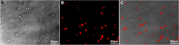
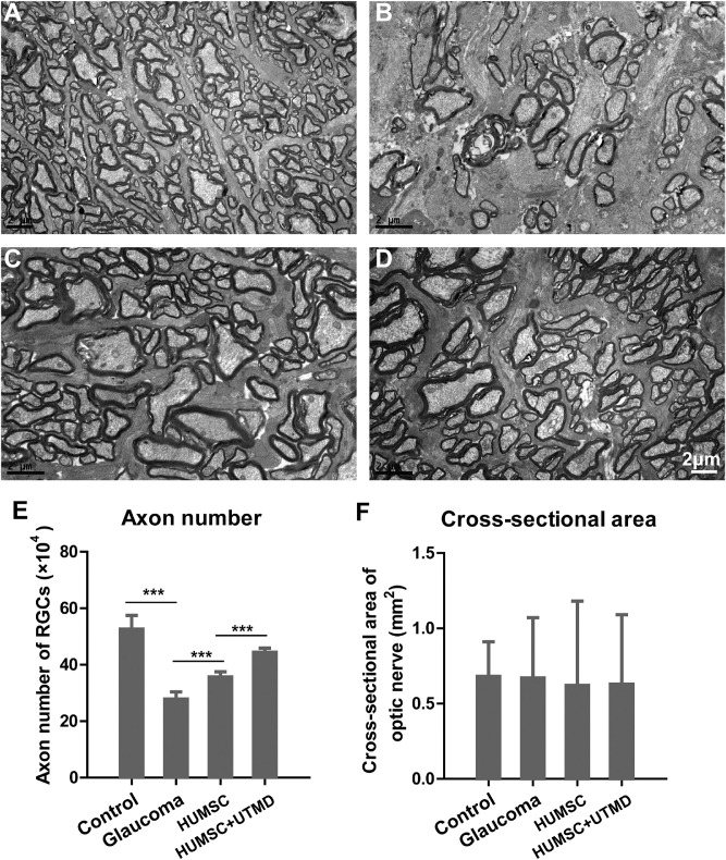
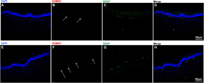
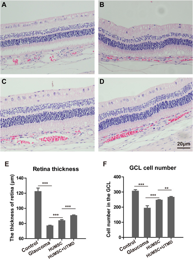
Eight New Zealand white healthy rabbits were used as the control group (group A). Twenty-four experimental glaucomatous rabbits were established as described previously and randomly divided into three groups: (1) received no treatment (group B); (2) received intravitreal transplantation of HUMSCs (group C); and (3) received UTMD-assisted intravitreal transplantation of HUMSCs (group D). After 4 weeks of treatment, the distribution of HUMSCs, retinal thickness, layer structure, retinal ganglion cells (RGCs), and their axons were examined.
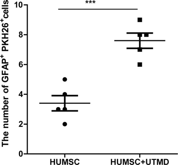
After 4 weeks of treatment, HUMSCs were successfully scattered under the retina. HUMSC transplantation significantly increased the regeneration of RGCs and their axons, and restored the retinal structure in glaucomatous rabbits. Moreover, the application of UTMD enhances HUMSC distribution and achieved more significant therapeutic effect.
Intravitreal transplantation of HUMSCs effectively repaired glaucomatous optic nerve injury, and UTMD enhanced the successful delivery of HUMSCs into injured retina, promoting its therapeutic effects remarkably.
This study demonstrated that HUMSC transplantation repaired the glaucoma-caused nerve injury significantly and the combination of UTMD can augment the therapeutic effect further, which has important clinical guiding significance for the development of therapeutic strategies of glaucoma.
本研究旨在探讨人脐带间充质干细胞(HUMSC)单独移植或辅助超声靶向微泡破坏(UTMD)治疗实验性兔青光眼新型实用模型中视神经病变的疗效。
8 只新西兰白兔作为对照组(A 组)。24 只实验性青光眼兔按先前描述的方法建立,并随机分为 3 组:(1)未治疗(B 组);(2)玻璃体内注射 HUMSCs(C 组);(3)UTMD 辅助玻璃体内注射 HUMSCs(D 组)。治疗 4 周后,观察 HUMSC 的分布、视网膜厚度、层结构、视网膜神经节细胞(RGCs)及其轴突。
治疗 4 周后,HUMSC 成功分散在视网膜下。HUMSC 移植显著增加了 RGCs 及其轴突的再生,恢复了青光眼兔的视网膜结构。此外,UTMD 的应用增强了 HUMSC 的分布,达到了更显著的治疗效果。
玻璃体内注射 HUMSC 可有效修复青光眼性视神经损伤,UTMD 增强了 HUMSC 向损伤视网膜的有效传递,显著提高了治疗效果。
本研究表明 HUMSC 移植显著修复了青光眼引起的神经损伤,联合应用 UTMD 可进一步增强治疗效果,对青光眼治疗策略的发展具有重要的临床指导意义。