• 文献检索
  • 文档翻译
  • 深度研究
  • 学术资讯
  • Suppr Zotero 插件Zotero 插件
  • 邀请有礼
  • 套餐&价格
  • 历史记录
应用&插件
Suppr Zotero 插件Zotero 插件浏览器插件Mac 客户端Windows 客户端微信小程序
定价
高级版会员购买积分包购买API积分包
服务
文献检索文档翻译深度研究API 文档MCP 服务
关于我们
关于 Suppr公司介绍联系我们用户协议隐私条款
关注我们

Suppr 超能文献

核心技术专利:CN118964589B侵权必究
粤ICP备2023148730 号-1Suppr @ 2026

文献检索

告别复杂PubMed语法,用中文像聊天一样搜索,搜遍4000万医学文献。AI智能推荐,让科研检索更轻松。

立即免费搜索

文件翻译

保留排版,准确专业,支持PDF/Word/PPT等文件格式,支持 12+语言互译。

免费翻译文档

深度研究

AI帮你快速写综述,25分钟生成高质量综述,智能提取关键信息,辅助科研写作。

立即免费体验

窄带成像放大内镜时代黏膜破损的定义

Definition of Mucosal Breaks in the Era of Magnifying Endoscopy with Narrow-Band Imaging.

作者信息

Kikuchi Daisuke, Odagiri Hiroyuki, Hoshihara Yoshio, Ochiai Yorinari, Suzuki Yugo, Hayasaka Junnosuke, Tanaka Masami, Nomura Kosuke, Yamashita Satoshi, Matsui Akira, Iizuka Toshiro, Hoteya Shu

机构信息

Department of Gastroenterology, Toranomon Hospital, Japan.

出版信息

Gastroenterol Res Pract. 2022 May 13;2022:3952962. doi: 10.1155/2022/3952962. eCollection 2022.

DOI:10.1155/2022/3952962
PMID:35601238
原文链接:https://pmc.ncbi.nlm.nih.gov/articles/PMC9122728/
Abstract

BACKGROUND

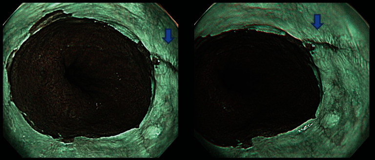
Gastroesophageal reflux disease is diagnosed endoscopically based on the presence of mucosal breaks. However, mucosal breaks can be judged differently depending on the endoscopist, even in the same image. We investigated how narrow-band imaging (NBI) and magnified endoscopy affect the judgment of mucosal breaks.

METHODS

A total of 43 consecutive patients were enrolled who had suspected mucosal breaks on white-light images (WLI) and underwent nonmagnified NBI (N-NBI) and magnified NBI (M-NBI) by a single endoscopist. From WLI, N-NBI, and M-NBI, 129 image files were created. Eight endoscopists reviewed the image files and judged the presence of mucosal breaks.

RESULTS

The 8 endoscopists determined mucosal breaks were present in 79.4 ± 9.5% (67.4%-93.0%) on WLI, and 76.7 ± 12.7% (53.5%-90.7%) on N-NBI. However, the percentage of mucosal breaks on M-NBI was significantly lower at 48.8 ± 17.0% (18.6%-65.1%) ( < 0.05). Intraclass correlation between observers was 0.864 (95% CI 0.793-0.918) for WLI and 0.863 (95% CI 0.791-0.917) for N-NBI but was lower for M-NBI at 0.758 (95% CI 0.631-0.854).

CONCLUSION

Rates of detection and agreement for mucosal breaks on WLI and N-NBI were high among endoscopists. However, these rates were lower on M-NBI.

摘要

背景

胃食管反流病通过内镜检查根据黏膜破损的存在来诊断。然而,即使在同一图像中,不同内镜医师对黏膜破损的判断也可能不同。我们研究了窄带成像(NBI)和放大内镜检查如何影响对黏膜破损的判断。

方法

连续纳入43例白光图像(WLI)上疑似有黏膜破损的患者,由一名内镜医师对其进行非放大NBI(N-NBI)和放大NBI(M-NBI)检查。从WLI、N-NBI和M-NBI中创建了129个图像文件。8名内镜医师对这些图像文件进行审查并判断是否存在黏膜破损。

结果

8名内镜医师判定WLI上黏膜破损的比例为79.4±9.5%(67.4%-93.),N-NBI上为76.7±12.7%(53.5%-90.7%)。然而,M-NBI上黏膜破损的比例显著较低,为48.8±17.0%(18.6%-65.1%)(P<0.05)。观察者之间的组内相关性,WLI为0.864(95%CI 0.793-0.918),N-NBI为0.863(95%CI 0.791-0.917),但M-NBI较低,为0.758(95%CI 0.631-0.854)。

结论

内镜医师对WLI和N-NBI上黏膜破损的检出率和一致性较高。然而,M-NBI上的这些率较低。

https://cdn.ncbi.nlm.nih.gov/pmc/blobs/7a4e/9122728/0d6141c91aa0/GRP2022-3952962.005.jpg
https://cdn.ncbi.nlm.nih.gov/pmc/blobs/7a4e/9122728/c060e2393d73/GRP2022-3952962.001.jpg
https://cdn.ncbi.nlm.nih.gov/pmc/blobs/7a4e/9122728/634a54711978/GRP2022-3952962.002.jpg
https://cdn.ncbi.nlm.nih.gov/pmc/blobs/7a4e/9122728/6fb88faf0b30/GRP2022-3952962.003.jpg
https://cdn.ncbi.nlm.nih.gov/pmc/blobs/7a4e/9122728/c91ca9901d77/GRP2022-3952962.004.jpg
https://cdn.ncbi.nlm.nih.gov/pmc/blobs/7a4e/9122728/0d6141c91aa0/GRP2022-3952962.005.jpg

相似文献

1
Definition of Mucosal Breaks in the Era of Magnifying Endoscopy with Narrow-Band Imaging.窄带成像放大内镜时代黏膜破损的定义
Gastroenterol Res Pract. 2022 May 13;2022:3952962. doi: 10.1155/2022/3952962. eCollection 2022.
2
Diagnostic performance of magnifying blue laser imaging versus magnifying narrow-band imaging for identifying the depth of invasion of superficial esophageal squamous cell carcinoma.放大蓝激光成像与放大窄带成像识别表浅性食管鳞状细胞癌浸润深度的诊断性能比较。
Dis Esophagus. 2021 Mar 8;34(3). doi: 10.1093/dote/doaa078.
3
Magnifying narrowband imaging is more accurate than conventional white-light imaging in diagnosis of gastric mucosal cancer.放大窄带成像比传统白光成像在诊断胃黏膜癌方面更准确。
Gastroenterology. 2011 Dec;141(6):2017-2025.e3. doi: 10.1053/j.gastro.2011.08.007. Epub 2011 Aug 19.
4
Comparative study on artificial intelligence systems for detecting early esophageal squamous cell carcinoma between narrow-band and white-light imaging.窄带成像与白光成像下人工智能系统检测早期食管鳞状细胞癌的对比研究
World J Gastroenterol. 2021 Jan 21;27(3):281-293. doi: 10.3748/wjg.v27.i3.281.
5
A comparative study of demarcation line diagnostic performance between non-magnifying observation with white light and non-magnifying observation with narrow-band light for early gastric cancer.非放大白光观察与非放大窄带光观察对早期胃癌的界限线诊断性能比较研究。
Gastric Cancer. 2022 Jul;25(4):761-769. doi: 10.1007/s10120-022-01299-5. Epub 2022 May 6.
6
Multicenter, prospective trial of white-light imaging alone versus white-light imaging followed by magnifying endoscopy with narrow-band imaging for the real-time imaging and diagnosis of invasion depth in superficial esophageal squamous cell carcinoma.多中心前瞻性试验:单纯白光成像与白光成像后联合窄带成像放大内镜用于浅表性食管鳞状细胞癌浸润深度的实时成像与诊断
Gastrointest Endosc. 2015;81(6):1355-1361.e2. doi: 10.1016/j.gie.2014.11.015. Epub 2015 Feb 12.
7
Is it proper to use non-magnified narrow-band imaging for esophageal neoplasia screening? Japanese single-center, prospective study.是否可以使用非放大窄带成像进行食管肿瘤筛查?日本单中心前瞻性研究。
Dig Endosc. 2012 Nov;24(6):412-8. doi: 10.1111/j.1443-1661.2012.01309.x. Epub 2012 Apr 10.
8
Assessment of novel endoscopic techniques for visualizing superficial esophageal squamous cell carcinoma: autofluorescence and narrow-band imaging.用于可视化浅表性食管鳞状细胞癌的新型内镜技术评估:自体荧光和窄带成像。
Dis Esophagus. 2009;22(5):439-46. doi: 10.1111/j.1442-2050.2008.00925.x. Epub 2009 Jan 23.
9
Texture and color enhancement imaging in magnifying endoscopic evaluation of colorectal adenomas.放大内镜评估大肠腺瘤中的纹理和颜色增强成像
World J Gastrointest Endosc. 2022 Feb 16;14(2):96-105. doi: 10.4253/wjge.v14.i2.96.
10
A training program of a new simplified classification of magnified narrow band imaging for superficial esophageal squamous cell carcinoma.一种新型放大窄带成像技术在食管浅表鳞癌分类中的培训方案。
J Gastroenterol Hepatol. 2018 Jun;33(6):1248-1255. doi: 10.1111/jgh.14071. Epub 2018 Mar 1.

引用本文的文献

1
Step-Down Therapy Using Vonoprazan for Giant Gastric Polyps in Long-Term Proton Pump Inhibitor Administration.在长期使用质子泵抑制剂治疗期间,使用沃克(富马酸伏诺拉生)进行降阶梯治疗巨大胃息肉。
Case Rep Gastroenterol. 2025 Mar 24;19(1):190-197. doi: 10.1159/000543876. eCollection 2025 Jan-Dec.
2
Efficacy of Step-Down Therapy Using Vonoprazan for Symptomatic Mild Reflux Esophagitis.使用沃克沙唑进行降阶梯治疗对有症状的轻度反流性食管炎的疗效。
Gastroenterol Res Pract. 2024 Nov 14;2024:5620034. doi: 10.1155/grp/5620034. eCollection 2024.

本文引用的文献

1
Statement for gastroesophageal reflux disease after peroral endoscopic myotomy from an international multicenter experience.经口内镜下肌切开术治疗胃食管反流病的国际多中心研究声明。
Esophagus. 2020 Jan;17(1):3-10. doi: 10.1007/s10388-019-00689-6. Epub 2019 Sep 26.
2
Interactions between Helicobacter pylori and gastroesophageal reflux disease.幽门螺杆菌与胃食管反流病之间的相互作用。
Esophagus. 2019 Jan;16(1):52-62. doi: 10.1007/s10388-018-0637-5. Epub 2018 Aug 27.
3
Utility of magnifying endoscopy with narrow band imaging in determining the invasion depth of superficial pharyngeal cancer.
窄带成像放大内镜在确定浅表性咽喉癌浸润深度中的应用
Head Neck. 2015 Jun;37(6):846-50. doi: 10.1002/hed.23683. Epub 2014 Jun 27.
4
Clinical feature of asymptomatic reflux esophagitis in patients who underwent upper gastrointestinal endoscopy.上消化道内镜检查患者无症状性反流性食管炎的临床特征。
J Gastroenterol Hepatol. 2012 Apr;27 Suppl 3:53-7. doi: 10.1111/j.1440-1746.2012.07073.x.
5
Magnifying narrow-band imaging versus magnifying white-light imaging for the differential diagnosis of gastric small depressive lesions: a prospective study.放大窄带成像与放大白光成像对胃小凹陷性病变的鉴别诊断:一项前瞻性研究。
Gastrointest Endosc. 2010 Mar;71(3):477-84. doi: 10.1016/j.gie.2009.10.036.
6
Early detection of superficial squamous cell carcinoma in the head and neck region and esophagus by narrow band imaging: a multicenter randomized controlled trial.窄带成像技术在头颈部和食管浅表鳞状细胞癌早期检测中的应用:一项多中心随机对照试验。
J Clin Oncol. 2010 Mar 20;28(9):1566-72. doi: 10.1200/JCO.2009.25.4680. Epub 2010 Feb 22.
7
A feasibility trial of narrow band imaging endoscopy in patients with gastroesophageal reflux disease.窄带成像内镜在胃食管反流病患者中的可行性试验。
Gastroenterology. 2007 Aug;133(2):454-64; quiz 674. doi: 10.1053/j.gastro.2007.06.006. Epub 2007 Jun 8.
8
Intraobserver and interobserver consistency for grading esophagitis with narrow-band imaging.使用窄带成像对食管炎进行分级的观察者内和观察者间一致性
Gastrointest Endosc. 2007 Aug;66(2):230-6. doi: 10.1016/j.gie.2006.10.056.
9
Clinical symptoms in endoscopic reflux esophagitis: evaluation in 8031 adult subjects.内镜下反流性食管炎的临床症状:8031例成年受试者的评估
Dig Dis Sci. 2003 Dec;48(12):2237-41. doi: 10.1023/b:ddas.0000007857.15694.15.
10
Comparison of inter- and intraobserver consistency for grading of esophagitis by expert and trainee endoscopists.专家内镜医师和实习内镜医师对食管炎分级的观察者间和观察者内一致性比较。
Gastrointest Endosc. 2002 Nov;56(5):639-43. doi: 10.1067/mge.2002.129220.