Liu Chin-Fu, Hsu Johnny, Xu Xin, Ramachandran Sandhya, Wang Victor, Miller Michael I, Hillis Argye E, Faria Andreia V
Center for Imaging Science, Johns Hopkins University, Baltimore, MD USA.
Department of Biomedical Engineering, Johns Hopkins University, Baltimore, MD USA.
Commun Med (Lond). 2021 Dec 16;1:61. doi: 10.1038/s43856-021-00062-8. eCollection 2021.
Accessible tools to efficiently detect and segment diffusion abnormalities in acute strokes are highly anticipated by the clinical and research communities.
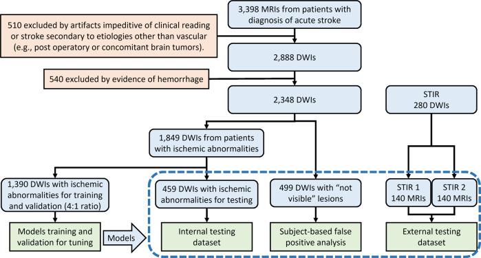
We developed a tool with deep learning networks trained and tested on a large dataset of 2,348 clinical diffusion weighted MRIs of patients with acute and sub-acute ischemic strokes, and further tested for generalization on 280 MRIs of an external dataset (STIR).
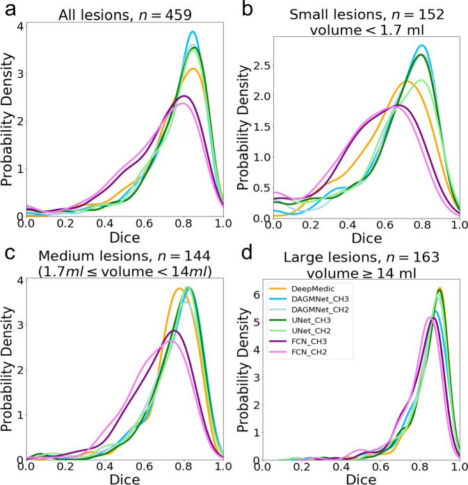
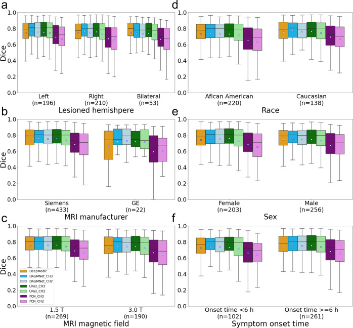
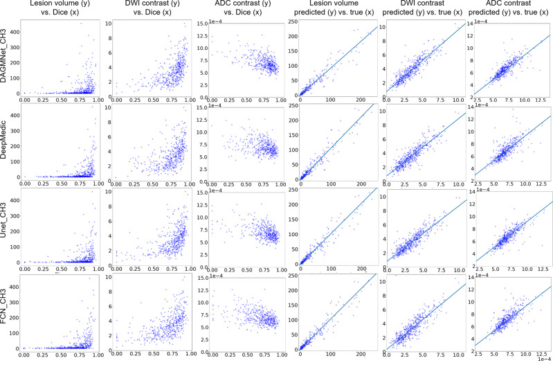
Our proposed model outperforms generic networks and DeepMedic, particularly in small lesions, with lower false positive rate, balanced precision and sensitivity, and robustness to data perturbs (e.g., artefacts, low resolution, technical heterogeneity). The agreement with human delineation rivals the inter-evaluator agreement; the automated lesion quantification of volume and contrast has virtually total agreement with human quantification.
Our tool is fast, public, accessible to non-experts, with minimal computational requirements, to detect and segment lesions via a single command line. Therefore, it fulfills the conditions to perform large scale, reliable and reproducible clinical and translational research.
临床和研究界迫切期待能有可便捷使用的工具,用于高效检测和分割急性中风中的扩散异常。
我们开发了一种工具,其深度学习网络在包含2348例急性和亚急性缺血性中风患者临床扩散加权磁共振成像的大型数据集上进行训练和测试,并在外部数据集(短TI反转恢复序列成像,STIR)的280例磁共振成像上进一步测试其泛化能力。
我们提出的模型优于通用网络和DeepMedic,尤其是在检测小病灶方面,具有更低的假阳性率、平衡的精度和灵敏度,以及对数据干扰(如伪影、低分辨率、技术异质性)的鲁棒性。与人工勾勒的一致性可媲美评估者间的一致性;自动进行的病灶体积和对比度量化与人工量化几乎完全一致。
我们的工具速度快且公开,非专业人员也可使用,计算要求极低,通过单一命令行即可检测和分割病灶。因此,它满足了开展大规模、可靠且可重复的临床和转化研究的条件。