O'Brien Hugh, Williams Michelle C, Rajani Ronak, Niederer Steven
School of Biomedical Engineering and Imaging Sciences, King's College London, London, United Kingdom.
Centre for Cardiovascular Science, University of Edinburgh, Edinburgh, United Kingdom.
Front Cardiovasc Med. 2022 May 12;9:847825. doi: 10.3389/fcvm.2022.847825. eCollection 2022.
Delayed enhancement CT (CT-DE) has been evaluated as a tool for the detection of myocardial scar and compares well to the gold standard of MRI with late gadolinium enhancement (MRI-LGE). Prior work has established that high performance can be achieved with manual reading; however, few studies have looked at quantitative measures to differentiate scar and healthy myocardium on CT-DE or automated analysis.
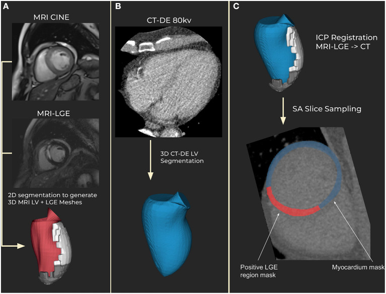
Eighteen patients with clinically indicated MRI-LGE were recruited for CT-DE at multiple 80 and 100 kV post contrast imaging. Left ventricle segmentation was performed on both imaging modalities, along with scar segmentation on MRI-LGE. Segmentations were registered together and scar regions were estimated on CT-DE. 93 radiomic features were calculated and analysed for their ability to differentiate between scarred and non-scarred myocardium regions. Machine learning (ML) classifiers were trained using the strongest set of radiomic features to classify segments containing scar on CT-DE. Features and classifiers were compared across both tube voltages and combined-energy images.
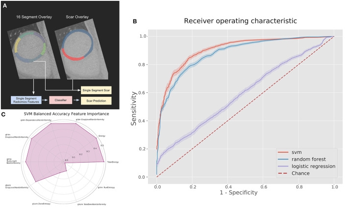
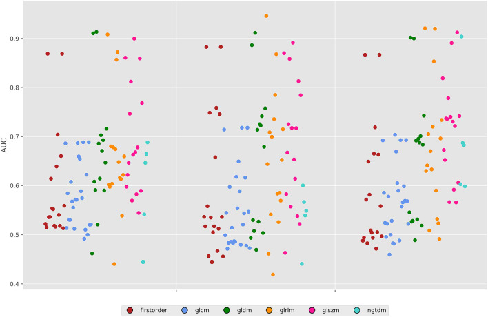
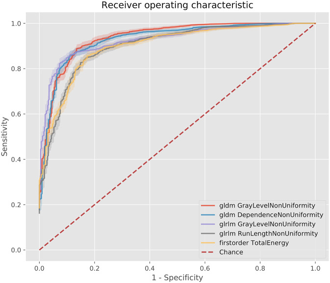
There were 59 and 51 statistically significant features in the 80 and 100 kV images respectively. Combined-energy imaging increased this to 63 with more features having area under the curve (AUC) above 0.9. The 10 highest AUC features for each image were used in the ML classifiers. The 100 kV images produced the best ML classifier, a support vector machine with an AUC of 0.88 (95% CI 0.87-0.90). Comparable performance was achieved with both the 80 kV and combined-energy images.
CT-DE can be quantitatively analyzed using radiomic feature calculations. These features may be suitable for ML classification techniques to prospectively identify AHA segments with performance comparable to previously reported manual reading. Future work on larger CT-DE datasets is warranted to establish optimum imaging parameters and features.
延迟增强CT(CT-DE)已被评估为检测心肌瘢痕的一种工具,并且与钆延迟增强磁共振成像(MRI-LGE)这一金标准相比表现良好。先前的研究已经证实,通过人工阅片可实现高性能;然而,很少有研究探讨在CT-DE上区分瘢痕和健康心肌的定量方法或自动分析。
招募18例有临床指征需进行MRI-LGE检查的患者,在造影后分别于80 kV和100 kV进行多次CT-DE成像。对两种成像方式均进行左心室分割,同时对MRI-LGE进行瘢痕分割。将分割结果进行配准,并在CT-DE上估计瘢痕区域。计算93个放射组学特征,并分析其区分瘢痕心肌区域和非瘢痕心肌区域的能力。使用最强的放射组学特征集训练机器学习(ML)分类器,以对CT-DE上包含瘢痕的节段进行分类。对两种管电压图像和能量合并图像的特征及分类器进行比较。
80 kV图像和100 kV图像分别有59个和51个具有统计学意义的特征。能量合并成像使这一数量增加到63个,更多特征的曲线下面积(AUC)高于0.9。ML分类器使用了每个图像中AUC最高的10个特征。100 kV图像产生了最佳的ML分类器,即支持向量机,其AUC为0.88(95%CI 0.87-0.90)。80 kV图像和能量合并图像均取得了可比的性能。
CT-DE可通过放射组学特征计算进行定量分析。这些特征可能适用于ML分类技术,以前瞻性地识别美国心脏协会(AHA)节段,其性能与先前报道的人工阅片相当。有必要对更大的CT-DE数据集开展进一步研究,以确定最佳成像参数和特征。