Baeßler Bettina, Götz Michael, Antoniades Charalambos, Heidenreich Julius F, Leiner Tim, Beer Meinrad
Department of Diagnostic and Interventional Radiology, University Hospital Würzburg, Würzburg, Germany.
Division of Experimental Radiology, Department for Diagnostic and Interventional Radiology, University Hospital Ulm, Ulm, Germany.
Front Cardiovasc Med. 2023 Feb 16;10:1120361. doi: 10.3389/fcvm.2023.1120361. eCollection 2023.
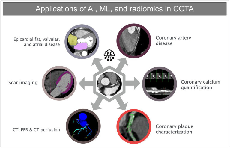
Coronary computed tomography angiography (CCTA) is increasingly the cornerstone in the management of patients with chronic coronary syndromes. This fact is reflected by current guidelines, which show a fundamental shift towards non-invasive imaging - especially CCTA. The guidelines for acute and stable coronary artery disease (CAD) of the European Society of Cardiology from 2019 and 2020 emphasize this shift. However, to fulfill this new role, a broader availability in adjunct with increased robustness of data acquisition and speed of data reporting of CCTA is needed. Artificial intelligence (AI) has made enormous progress for all imaging methodologies concerning (semi)-automatic tools for data acquisition and data post-processing, with outreach toward decision support systems. Besides onco- and neuroimaging, cardiac imaging is one of the main areas of application. Most current AI developments in the scenario of cardiac imaging are related to data postprocessing. However, AI applications (including radiomics) for CCTA also should enclose data acquisition (especially the fact of dose reduction) and data interpretation (presence and extent of CAD). The main effort will be to integrate these AI-driven processes into the clinical workflow, and to combine imaging data/results with further clinical data, thus - beyond the diagnosis of CAD- enabling prediction and forecast of morbidity and mortality. Furthermore, data fusing for therapy planning (e.g., invasive angiography/TAVI planning) will be warranted. The aim of this review is to present a holistic overview of AI applications in CCTA (including radiomics) under the umbrella of clinical workflows and clinical decision-making. The review first summarizes and analyzes applications for the main role of CCTA, i.e., to non-invasively rule out stable coronary artery disease. In the second step, AI applications for additional diagnostic purposes, i.e., to improve diagnostic power (CAC = coronary artery classifications), improve differential diagnosis (CT-FFR and CT perfusion), and finally improve prognosis (again CAC plus epi- and pericardial fat analysis) are reviewed.
冠状动脉计算机断层扫描血管造影(CCTA)日益成为慢性冠状动脉综合征患者管理的基石。这一事实在当前指南中得到了体现,这些指南显示出向非侵入性成像——尤其是CCTA的根本转变。欧洲心脏病学会2019年和2020年的急性和稳定冠状动脉疾病(CAD)指南强调了这一转变。然而,为了履行这一新角色,需要更广泛地提供CCTA,并提高数据采集的稳健性和数据报告的速度。人工智能(AI)在所有成像方法的(半)自动数据采集和数据后处理工具方面取得了巨大进展,并向决策支持系统拓展。除了肿瘤和神经成像外,心脏成像是主要应用领域之一。目前心脏成像领域的大多数人工智能发展都与数据后处理有关。然而,CCTA的人工智能应用(包括放射组学)也应涵盖数据采集(尤其是剂量减少这一事实)和数据解读(CAD的存在和范围)。主要努力将是把这些人工智能驱动的过程整合到临床工作流程中,并将成像数据/结果与进一步的临床数据相结合,从而——除了CAD诊断之外——实现发病率和死亡率的预测和预报。此外,还将有必要进行用于治疗规划(例如,侵入性血管造影/TAVI规划)的数据融合。本综述的目的是在临床工作流程和临床决策的框架下,对CCTA中的人工智能应用(包括放射组学)进行全面概述。该综述首先总结并分析了CCTA主要作用的应用,即无创排除稳定冠状动脉疾病。第二步,综述了用于其他诊断目的的人工智能应用,即提高诊断能力(CAC = 冠状动脉分类)、改善鉴别诊断(CT-FFR和CT灌注),最后改善预后(同样是CAC加上心外膜和心包脂肪分析)。