Institute of Statistical Science, Academia Sinica, Taipei, Taiwan.
Department of Computer Science, National Tsing Hua University, Hsinchu, Taiwan.
Brain Topogr. 2022 Jul;35(4):375-397. doi: 10.1007/s10548-022-00897-x. Epub 2022 Jun 6.
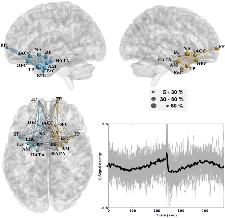
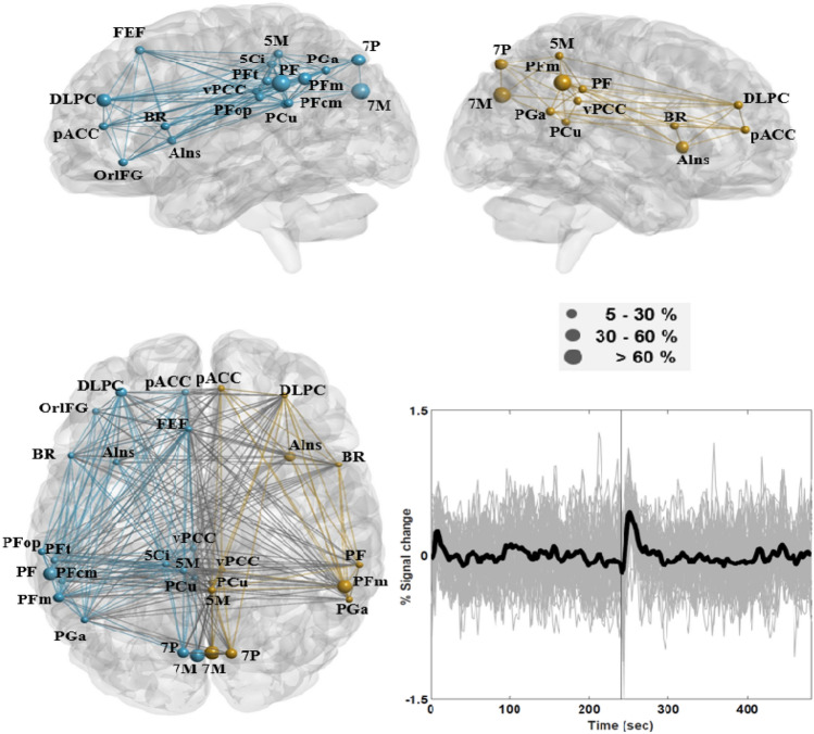
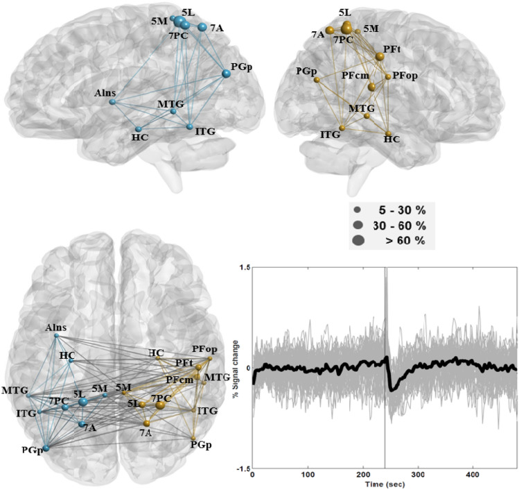
This study empirically assessed the strength and duration of short-term effects induced by brain reactions to closing/opening the eyes on a few well-known resting-state networks. We also examined the association between these reactions and subjects' cortisol levels. A total of 55 young adults underwent 8-min resting-state fMRI (rs-fMRI) scans under 4-min eyes-closed and 4-min eyes-open conditions. Saliva samples were collected from 25 of the 55 subjects before and after the fMRI sessions and assayed for cortisol levels. Our empirical results indicate that when the subjects were relaxed with their eyes closed, the effect of opening the eyes on conventional resting-state networks (e.g., default-mode, frontal-parietal, and saliency networks) lasted for roughly 60-s, during which we observed a short-term increase in activity in rs-fMRI time courses. Moreover, brain reactions to opening the eyes had a pronounced effect on time courses in the temporo-parietal lobes and limbic structures, both of which presented a prolonged decrease in activity. After controlling for demographic factors, we observed a significantly positive correlation between pre-scan cortisol levels and connectivity in the limbic structures under both conditions. Under the eyes-closed condition, the temporo-parietal lobes presented significant connectivity to limbic structures and a significantly positive correlation with pre-scan cortisol levels. Future research on rs-fMRI could consider the eyes-closed condition when probing resting-state connectivity and its neuroendocrine correlates, such as cortisol levels. It also appears that abrupt instructions to open the eyes while the subject is resting quietly with eyes closed could be used to probe brain reactivity to aversive stimuli in the ventral hippocampus and other limbic structures.
本研究实证评估了大脑对闭眼/睁眼反应在几个著名静息态网络上产生的短期效应的强度和持续时间。我们还研究了这些反应与被试皮质醇水平之间的关联。总共 55 名年轻成年人在闭眼 4 分钟和睁眼 4 分钟的条件下进行了 8 分钟的静息态 fMRI(rs-fMRI)扫描。从 55 名受试者中的 25 名采集了 fMRI 前后的唾液样本,并检测了皮质醇水平。我们的实验结果表明,当被试者闭眼放松时,睁眼对传统静息态网络(如默认模式、额顶叶和突显网络)的影响持续约 60 秒,在此期间我们观察到 rs-fMRI 时间序列中短期活动增加。此外,大脑对睁眼的反应对颞顶叶和边缘结构的时间序列有明显影响,两者的活动均呈现出长时间的下降。在控制了人口统计学因素后,我们观察到在两种条件下,皮质醇水平与边缘结构的连接之间存在显著的正相关。在闭眼条件下,颞顶叶与边缘结构之间呈现出显著的连接,与皮质醇水平的相关性呈显著正相关。未来的 rs-fMRI 研究可以考虑在探测静息态连接及其神经内分泌相关性(如皮质醇水平)时采用闭眼条件。此外,在被试者安静闭眼休息时突然指令睁眼,可能会被用来探测腹侧海马体和其他边缘结构中对厌恶刺激的大脑反应。