Department of Physiology, University of Maryland School of Medicine, Baltimore, MD 21201.
Program in Neuroscience, University of Maryland School of Medicine, Baltimore, MD 21201.
Proc Natl Acad Sci U S A. 2024 Apr 23;121(17):e2315379121. doi: 10.1073/pnas.2315379121. Epub 2024 Apr 16.
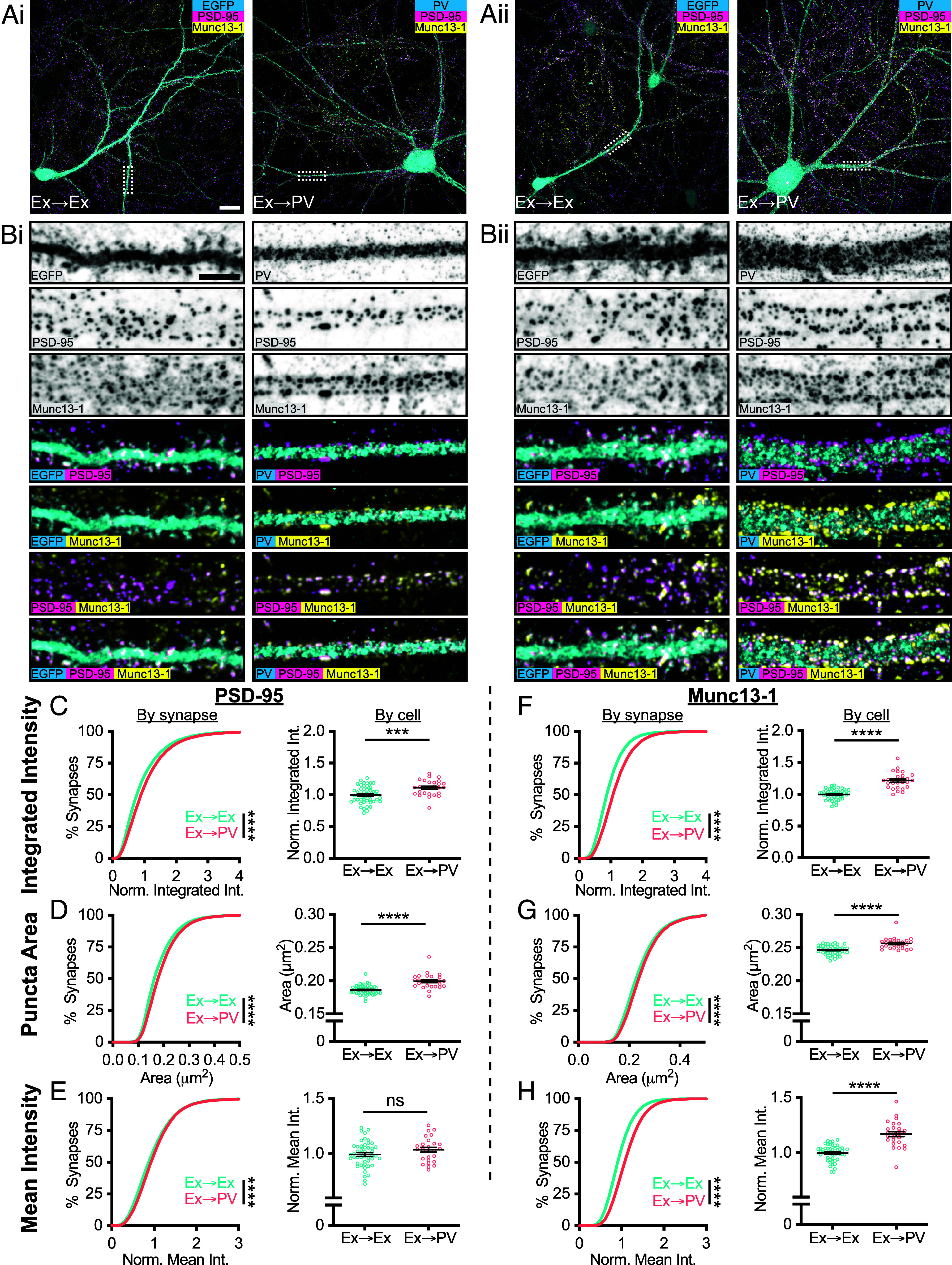
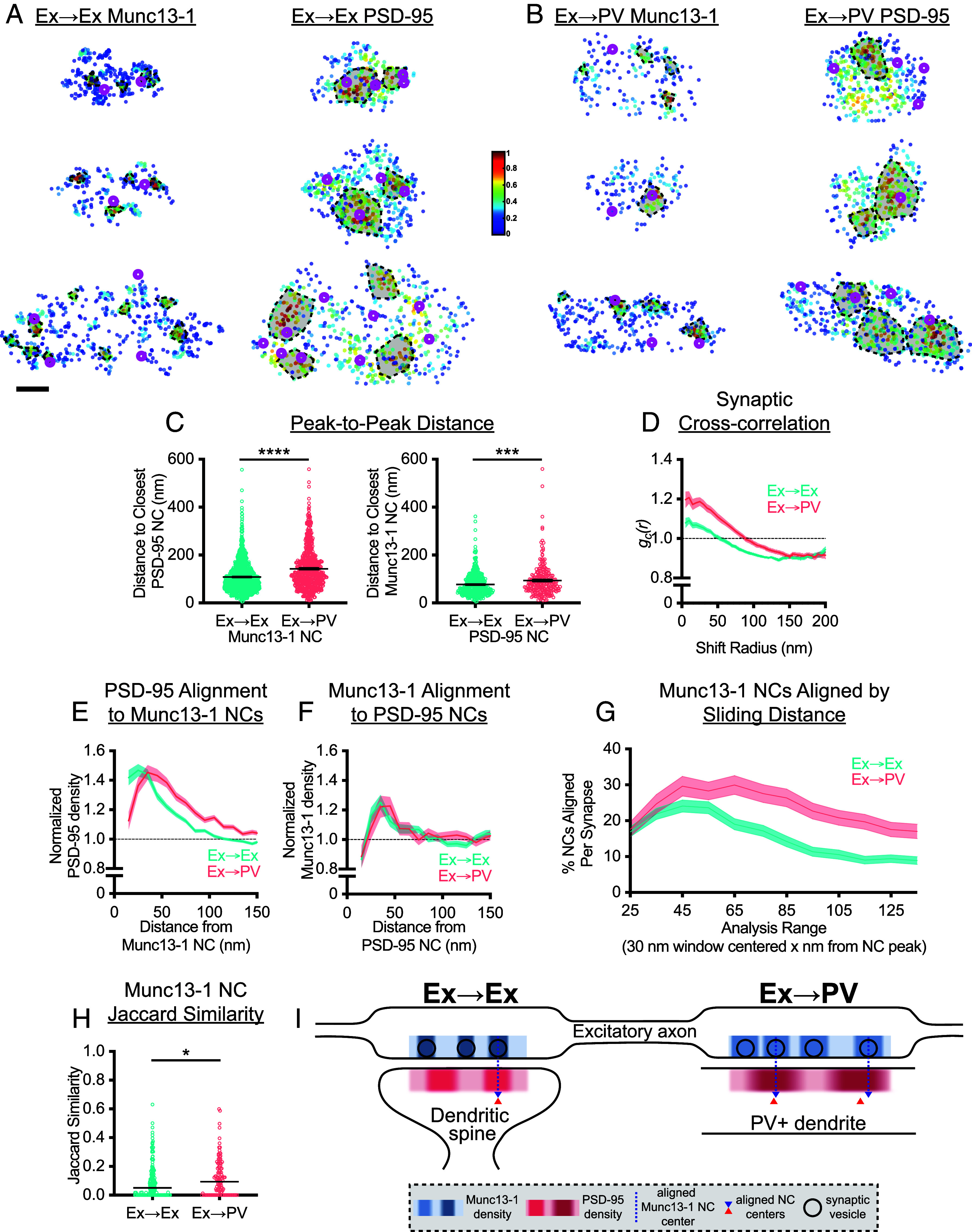
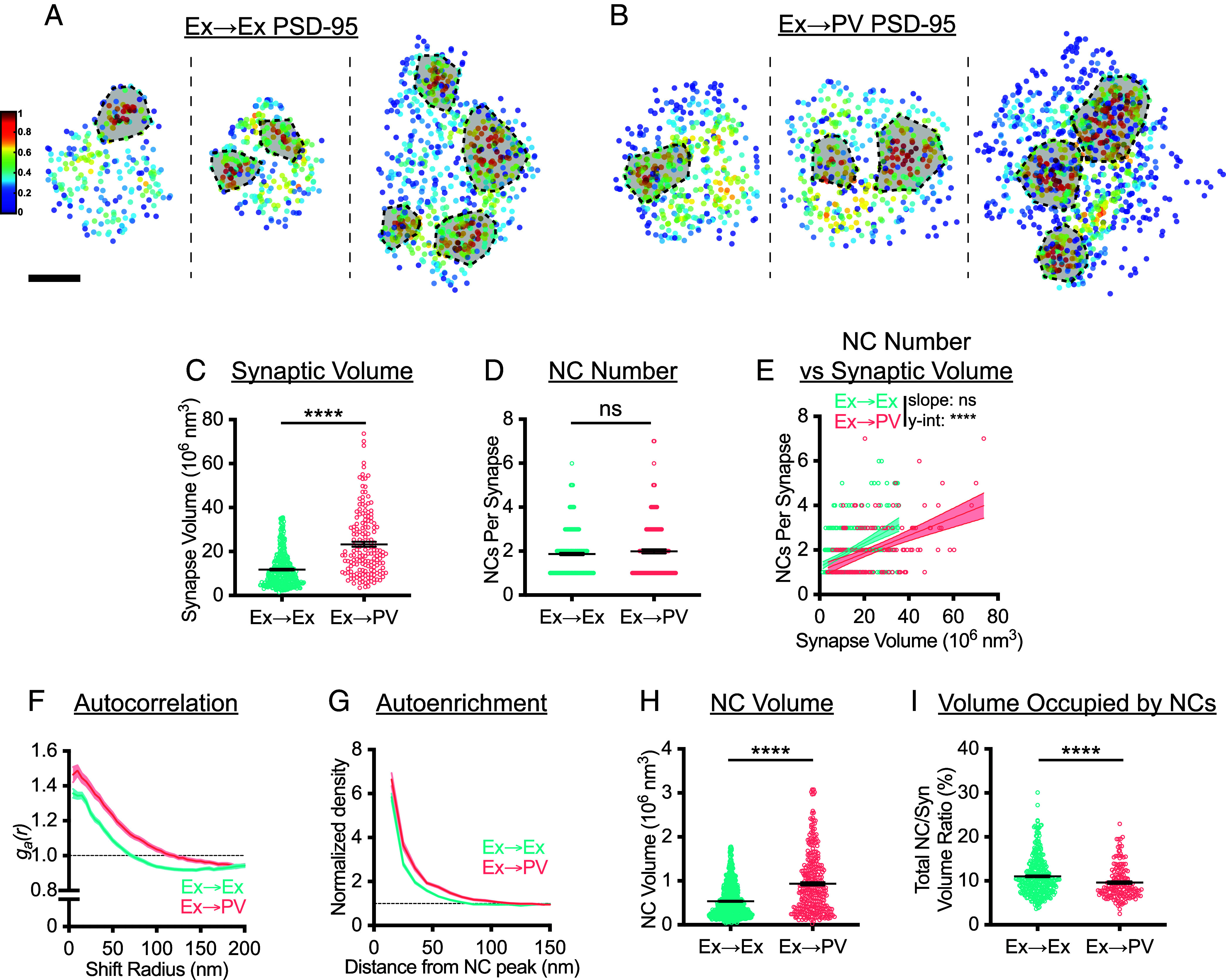
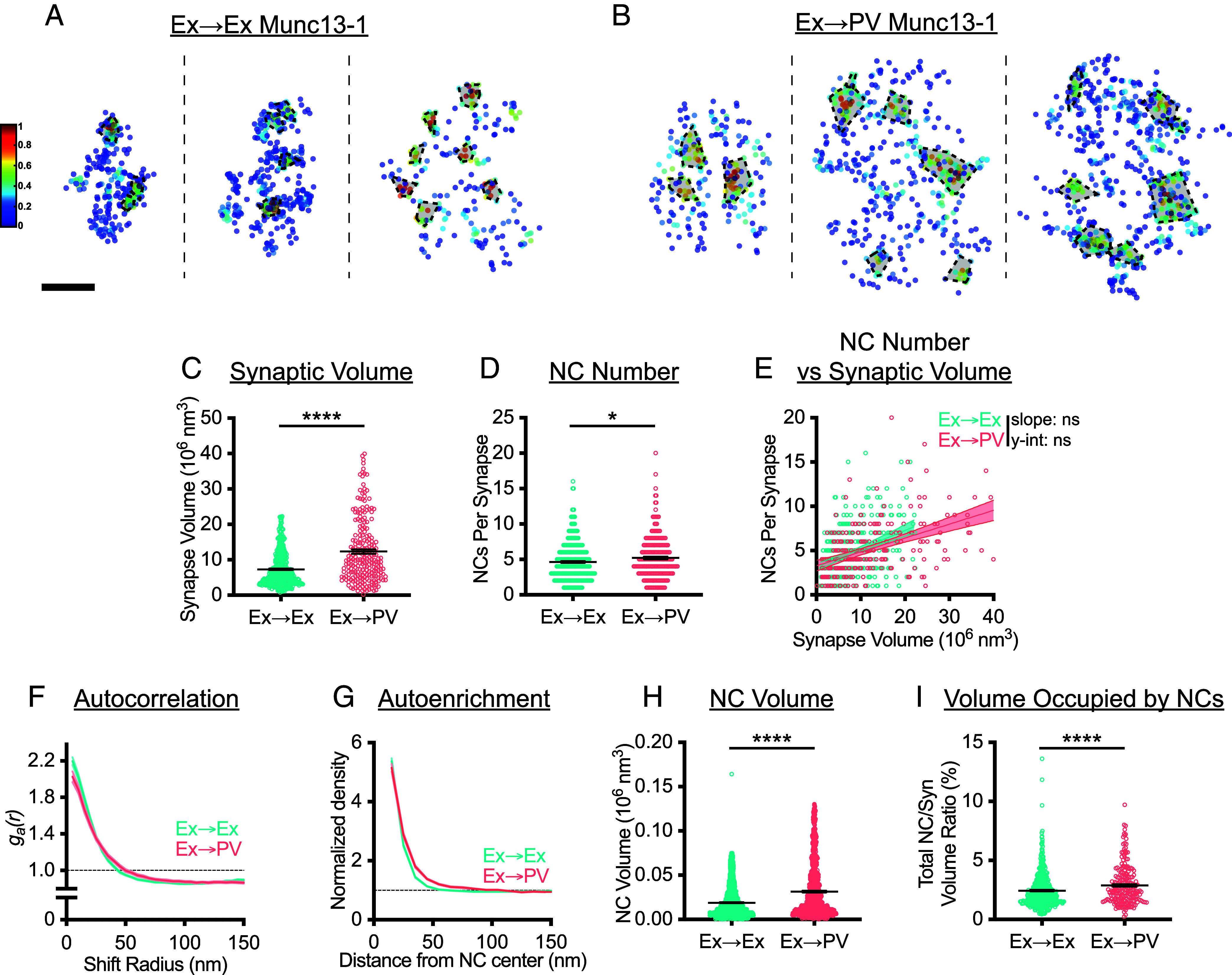
A key feature of excitatory synapses is the existence of subsynaptic protein nanoclusters (NCs) whose precise alignment across the cleft in a transsynaptic nanocolumn influences the strength of synaptic transmission. However, whether nanocolumn properties vary between excitatory synapses functioning in different cellular contexts is unknown. We used a combination of confocal and DNA-PAINT super-resolution microscopy to directly compare the organization of shared scaffold proteins at two important excitatory synapses-those forming onto excitatory principal neurons (Ex→Ex synapses) and those forming onto parvalbumin-expressing interneurons (Ex→PV synapses). As in Ex→Ex synapses, we find that in Ex→PV synapses, presynaptic Munc13-1 and postsynaptic PSD-95 both form NCs that demonstrate alignment, underscoring synaptic nanostructure and the transsynaptic nanocolumn as conserved organizational principles of excitatory synapses. Despite the general conservation of these features, we observed specific differences in the characteristics of pre- and postsynaptic Ex→PV nanostructure. Ex→PV synapses contained larger PSDs with fewer PSD-95 NCs when accounting for size than Ex→Ex synapses. Furthermore, the PSD-95 NCs were larger and denser. The identity of the postsynaptic cell was also represented in Munc13-1 organization, as Ex→PV synapses hosted larger Munc13-1 puncta that contained less dense but larger and more numerous Munc13-1 NCs. Moreover, we measured the spatial variability of transsynaptic alignment in these synapse types, revealing protein alignment in Ex→PV synapses over a distinct range of distances compared to Ex→Ex synapses. We conclude that while general principles of nanostructure and alignment are shared, cell-specific elements of nanodomain organization likely contribute to functional diversity of excitatory synapses.
兴奋性突触的一个关键特征是存在亚突触蛋白纳米簇(NCs),其在跨突触纳米柱中的裂谷中精确排列会影响突触传递的强度。然而,在不同细胞环境中发挥作用的兴奋性突触之间的纳米柱特性是否存在差异尚不清楚。我们使用共聚焦和 DNA-PAINT 超分辨率显微镜的组合,直接比较了两种重要的兴奋性突触(形成于兴奋性主神经元上的突触[Ex→Ex 突触]和形成于表达 Parvalbumin 的中间神经元上的突触[Ex→PV 突触])中共享支架蛋白的组织。与 Ex→Ex 突触一样,我们发现,在 Ex→PV 突触中,突触前 Munc13-1 和突触后 PSD-95 都形成了表现出对齐的 NC,这突出了突触纳米结构和跨突触纳米柱作为兴奋性突触的保守组织原则。尽管这些特征具有普遍的保守性,但我们观察到前突触和后突触 Ex→PV 纳米结构的特征存在特定差异。与 Ex→Ex 突触相比,当考虑到大小时,Ex→PV 突触的 PSD 较大,PSD-95 NC 较少。此外,PSD-95 NC 较大且较密。后突触细胞的身份也反映在 Munc13-1 的组织中,因为 Ex→PV 突触拥有较大的 Munc13-1 斑点,其包含较不密集但较大和更多的 Munc13-1 NC。此外,我们测量了这些突触类型中转突触对齐的空间变异性,揭示了 Ex→PV 突触中的蛋白质对齐在与 Ex→Ex 突触相比的明显距离范围内。我们得出的结论是,虽然纳米结构和对齐的一般原则是共享的,但纳米结构域组织的细胞特异性元素可能有助于兴奋性突触的功能多样性。