• 文献检索
  • 文档翻译
  • 深度研究
  • 学术资讯
  • Suppr Zotero 插件Zotero 插件
  • 邀请有礼
  • 套餐&价格
  • 历史记录
应用&插件
Suppr Zotero 插件Zotero 插件浏览器插件Mac 客户端Windows 客户端微信小程序
定价
高级版会员购买积分包购买API积分包
服务
文献检索文档翻译深度研究API 文档MCP 服务
关于我们
关于 Suppr公司介绍联系我们用户协议隐私条款
关注我们

Suppr 超能文献

核心技术专利:CN118964589B侵权必究
粤ICP备2023148730 号-1Suppr @ 2026

文献检索

告别复杂PubMed语法,用中文像聊天一样搜索,搜遍4000万医学文献。AI智能推荐,让科研检索更轻松。

立即免费搜索

文件翻译

保留排版,准确专业,支持PDF/Word/PPT等文件格式,支持 12+语言互译。

免费翻译文档

深度研究

AI帮你快速写综述,25分钟生成高质量综述,智能提取关键信息,辅助科研写作。

立即免费体验

使用卷积神经网络识别左右光学相干断层扫描图像之间的不对称性。

Asymmetry between right and left optical coherence tomography images identified using convolutional neural networks.

机构信息

Department of Ophthalmology, Gyeongsang National University Changwon Hospital, #11 Samjeongja-ro, Seongsan-gu, Changwon, 51472, Republic of Korea.

Department of Ophthalmology, Gyeongsang National University College of Medicine, Institute of Health Sciences, Jinju, Republic of Korea.

出版信息

Sci Rep. 2022 Jun 15;12(1):9925. doi: 10.1038/s41598-022-14140-x.

DOI:10.1038/s41598-022-14140-x
PMID:35705663
原文链接:https://pmc.ncbi.nlm.nih.gov/articles/PMC9200978/
Abstract

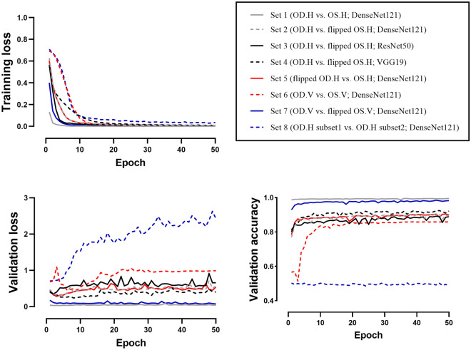
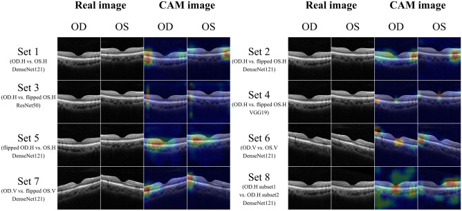
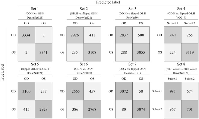
In a previous study, we identified biocular asymmetries in fundus photographs, and macula was discriminative area to distinguish left and right fundus images with > 99.9% accuracy. The purposes of this study were to investigate whether optical coherence tomography (OCT) images of the left and right eyes could be discriminated by convolutional neural networks (CNNs) and to support the previous result. We used a total of 129,546 OCT images. CNNs identified right and left horizontal images with high accuracy (99.50%). Even after flipping the left images, all of the CNNs were capable of discriminating them (DenseNet121: 90.33%, ResNet50: 88.20%, VGG19: 92.68%). The classification accuracy results were similar for the right and left flipped images (90.24% vs. 90.33%, respectively; p = 0.756). The CNNs also differentiated right and left vertical images (86.57%). In all cases, the discriminatory ability of the CNNs yielded a significant p value (< 0.001). However, the CNNs could not well-discriminate right horizontal images (50.82%, p = 0.548). There was a significant difference in identification accuracy between right and left horizontal and vertical OCT images and between flipped and non-flipped images. As this could result in bias in machine learning, care should be taken when flipping images.

摘要

在之前的研究中,我们发现眼底照片存在双眼不对称性,而黄斑是区分左右眼底图像的有区分性区域,准确率超过 99.9%。本研究旨在探讨卷积神经网络(CNN)是否可以区分左右眼的光学相干断层扫描(OCT)图像,并验证之前的结果。我们共使用了 129546 张 OCT 图像。CNN 能够非常准确地识别左右水平图像(99.50%)。即使对左眼图像进行翻转,所有 CNN 都能够对其进行区分(DenseNet121:90.33%,ResNet50:88.20%,VGG19:92.68%)。左右翻转图像的分类准确率结果相似(分别为 90.24%和 90.33%,p=0.756)。CNN 还可以区分左右垂直图像(86.57%)。在所有情况下,CNN 的区分能力都产生了显著的 p 值(<0.001)。然而,CNN 无法很好地区分右眼水平图像(50.82%,p=0.548)。左右水平和垂直 OCT 图像以及翻转和非翻转图像之间的识别准确率存在显著差异。由于这可能导致机器学习中的偏差,因此在翻转图像时应谨慎。

https://cdn.ncbi.nlm.nih.gov/pmc/blobs/0f5d/9200978/dd72c78000d4/41598_2022_14140_Fig5_HTML.jpg
https://cdn.ncbi.nlm.nih.gov/pmc/blobs/0f5d/9200978/7bd7fad2c849/41598_2022_14140_Fig1_HTML.jpg
https://cdn.ncbi.nlm.nih.gov/pmc/blobs/0f5d/9200978/3d369c17a829/41598_2022_14140_Fig2_HTML.jpg
https://cdn.ncbi.nlm.nih.gov/pmc/blobs/0f5d/9200978/6a57492d65d4/41598_2022_14140_Fig3_HTML.jpg
https://cdn.ncbi.nlm.nih.gov/pmc/blobs/0f5d/9200978/20fd4e514eb8/41598_2022_14140_Fig4_HTML.jpg
https://cdn.ncbi.nlm.nih.gov/pmc/blobs/0f5d/9200978/dd72c78000d4/41598_2022_14140_Fig5_HTML.jpg

相似文献

1
Asymmetry between right and left optical coherence tomography images identified using convolutional neural networks.使用卷积神经网络识别左右光学相干断层扫描图像之间的不对称性。
Sci Rep. 2022 Jun 15;12(1):9925. doi: 10.1038/s41598-022-14140-x.
2
Asymmetry between right and left fundus images identified using convolutional neural networks.使用卷积神经网络识别眼底左右图像的不对称性。
Sci Rep. 2022 Jan 27;12(1):1444. doi: 10.1038/s41598-021-04323-3.
3
The possibility of the combination of OCT and fundus images for improving the diagnostic accuracy of deep learning for age-related macular degeneration: a preliminary experiment.OCT 与眼底图像相结合提高深度学习诊断年龄相关性黄斑变性准确性的可能性:一项初步实验。
Med Biol Eng Comput. 2019 Mar;57(3):677-687. doi: 10.1007/s11517-018-1915-z. Epub 2018 Oct 22.
4
Automated diagnoses of age-related macular degeneration and polypoidal choroidal vasculopathy using bi-modal deep convolutional neural networks.使用双模态深度卷积神经网络进行与年龄相关的黄斑变性和息肉样脉络膜血管病变的自动诊断。
Br J Ophthalmol. 2021 Apr;105(4):561-566. doi: 10.1136/bjophthalmol-2020-315817. Epub 2020 Jun 4.
5
Classification of pachychoroid on optical coherence tomography using deep learning.利用深度学习对光学相干断层扫描图像中的脉络膜增厚进行分类。
Graefes Arch Clin Exp Ophthalmol. 2021 Jul;259(7):1803-1809. doi: 10.1007/s00417-021-05104-4. Epub 2021 Feb 22.
6
Calibration of fundus images using spectral domain optical coherence tomography.使用光谱域光学相干断层扫描对眼底图像进行校准。
Ophthalmic Surg Lasers Imaging. 2008 Jul-Aug;39(4 Suppl):S15-20. doi: 10.3928/15428877-20080715-01.
7
Automated detection of exudative age-related macular degeneration in spectral domain optical coherence tomography using deep learning.使用深度学习在光谱域光学相干断层扫描中自动检测渗出性年龄相关性黄斑变性。
Graefes Arch Clin Exp Ophthalmol. 2018 Feb;256(2):259-265. doi: 10.1007/s00417-017-3850-3. Epub 2017 Nov 20.
8
Two- and three-dimensional topographic analysis of pathologically myopic eyes with dome-shaped macula and inferior staphyloma by spectral domain optical coherence tomography.使用光谱域光学相干断层扫描对患有穹窿状黄斑和下方葡萄肿的病理性近视眼睛进行二维和三维地形分析。
Graefes Arch Clin Exp Ophthalmol. 2017 May;255(5):903-912. doi: 10.1007/s00417-017-3587-z. Epub 2017 Jan 17.
9
Automatic detection of retinal regions using fully convolutional networks for diagnosis of abnormal maculae in optical coherence tomography images.利用全卷积网络自动检测视网膜区域,用于光学相干断层扫描图像中黄斑异常的诊断。
J Biomed Opt. 2019 May;24(5):1-9. doi: 10.1117/1.JBO.24.5.056003.
10
A Hybrid Model Composed of Two Convolutional Neural Networks (CNNs) for Automatic Retinal Layer Segmentation of OCT Images in Retinitis Pigmentosa (RP).一种由两个卷积神经网络(CNNs)组成的混合模型,用于自动分割色素性视网膜炎(RP)的 OCT 图像中的视网膜层。
Transl Vis Sci Technol. 2021 Nov 1;10(13):9. doi: 10.1167/tvst.10.13.9.

引用本文的文献

1
A Low Complexity Efficient Deep Learning Model for Automated Retinal Disease Diagnosis.一种用于视网膜疾病自动诊断的低复杂度高效深度学习模型。
J Healthc Inform Res. 2025 Jan 3;9(1):1-40. doi: 10.1007/s41666-024-00182-5. eCollection 2025 Mar.
2
Exploring Publicly Accessible Optical Coherence Tomography Datasets: A Comprehensive Overview.探索可公开获取的光学相干断层扫描数据集:全面概述
Diagnostics (Basel). 2024 Aug 1;14(15):1668. doi: 10.3390/diagnostics14151668.
3
Application of deep learning artificial intelligence technique to the classification of clinical orthodontic photos.

本文引用的文献

1
Asymmetry between right and left fundus images identified using convolutional neural networks.使用卷积神经网络识别眼底左右图像的不对称性。
Sci Rep. 2022 Jan 27;12(1):1444. doi: 10.1038/s41598-021-04323-3.
2
Assessment of patient specific information in the wild on fundus photography and optical coherence tomography.眼底照相和光相干断层扫描的野生患者特定信息评估。
Sci Rep. 2021 Apr 21;11(1):8621. doi: 10.1038/s41598-021-86577-5.
3
Array programming with NumPy.使用 NumPy 进行数组编程。
深度学习人工智能技术在临床正畸照片分类中的应用。
BMC Oral Health. 2022 Oct 25;22(1):454. doi: 10.1186/s12903-022-02466-x.
Nature. 2020 Sep;585(7825):357-362. doi: 10.1038/s41586-020-2649-2. Epub 2020 Sep 16.
4
Peripapillary Retinal Nerve Fiber Layer Profile in Relation to Refractive Error and Axial Length: Results From the Gutenberg Health Study.视乳头周围视网膜神经纤维层形态与屈光不正和眼轴长度的关系:古登堡健康研究结果
Transl Vis Sci Technol. 2020 Aug 21;9(9):35. doi: 10.1167/tvst.9.9.35. eCollection 2020 Aug.
5
Effects of Hypertension, Diabetes, and Smoking on Age and Sex Prediction from Retinal Fundus Images.高血压、糖尿病和吸烟对视网膜眼底图像年龄和性别预测的影响。
Sci Rep. 2020 Mar 12;10(1):4623. doi: 10.1038/s41598-020-61519-9.
6
Convolutional Neural Networks as a Model of the Visual System: Past, Present, and Future.作为视觉系统模型的卷积神经网络:过去、现在与未来。
J Cogn Neurosci. 2021 Sep 1;33(10):2017-2031. doi: 10.1162/jocn_a_01544.
7
Referable diabetic retinopathy identification from eye fundus images with weighted path for convolutional neural network.基于卷积神经网络加权路径的眼底图像可归因糖尿病性视网膜病变识别。
Artif Intell Med. 2019 Aug;99:101694. doi: 10.1016/j.artmed.2019.07.002. Epub 2019 Jul 10.
8
Microaneurysm detection in fundus images using a two-step convolutional neural network.使用两步卷积神经网络检测眼底图像中的微动脉瘤。
Biomed Eng Online. 2019 May 29;18(1):67. doi: 10.1186/s12938-019-0675-9.
9
Artificial intelligence in retina.视网膜中的人工智能。
Prog Retin Eye Res. 2018 Nov;67:1-29. doi: 10.1016/j.preteyeres.2018.07.004. Epub 2018 Aug 1.
10
Convolutional neural networks: an overview and application in radiology.卷积神经网络:概述及其在放射学中的应用。
Insights Imaging. 2018 Aug;9(4):611-629. doi: 10.1007/s13244-018-0639-9. Epub 2018 Jun 22.