• 文献检索
  • 文档翻译
  • 深度研究
  • 学术资讯
  • Suppr Zotero 插件Zotero 插件
  • 邀请有礼
  • 套餐&价格
  • 历史记录
应用&插件
Suppr Zotero 插件Zotero 插件浏览器插件Mac 客户端Windows 客户端微信小程序
定价
高级版会员购买积分包购买API积分包
服务
文献检索文档翻译深度研究API 文档MCP 服务
关于我们
关于 Suppr公司介绍联系我们用户协议隐私条款
关注我们

Suppr 超能文献

核心技术专利:CN118964589B侵权必究
粤ICP备2023148730 号-1Suppr @ 2026

文献检索

告别复杂PubMed语法,用中文像聊天一样搜索,搜遍4000万医学文献。AI智能推荐,让科研检索更轻松。

立即免费搜索

文件翻译

保留排版,准确专业,支持PDF/Word/PPT等文件格式,支持 12+语言互译。

免费翻译文档

深度研究

AI帮你快速写综述,25分钟生成高质量综述,智能提取关键信息,辅助科研写作。

立即免费体验

基于增强CT影像组学列线图的1型和2型乳头状肾细胞癌的鉴别诊断

Differential Diagnosis of Type 1 and Type 2 Papillary Renal Cell Carcinoma Based on Enhanced CT Radiomics Nomogram.

作者信息

Gao Yankun, Wang Xingwei, Wang Shihui, Miao Yingying, Zhu Chao, Li Cuiping, Huang Guoquan, Jiang Yan, Li Jianying, Zhao Xiaoying, Wu Xingwang

机构信息

Department of Radiology, The First Affiliated Hospital of Anhui Medical University, Hefei, China.

Department of Radiology, The First Affiliated Hospital of Wannan Medical college, Wuhu, China.

出版信息

Front Oncol. 2022 Jun 3;12:854979. doi: 10.3389/fonc.2022.854979. eCollection 2022.

DOI:10.3389/fonc.2022.854979
PMID:35719928
原文链接:https://pmc.ncbi.nlm.nih.gov/articles/PMC9204229/
Abstract

OBJECTIVES

To construct a contrast-enhanced CT-based radiomics nomogram that combines clinical factors and a radiomics signature to distinguish papillary renal cell carcinoma (pRCC) type 1 from pRCC type 2 tumours.

METHODS

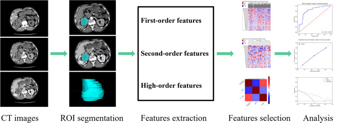
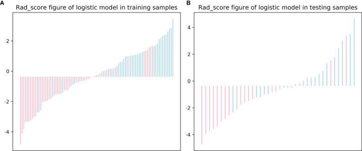
A total of 131 patients with 60 in pRCC type 1 and 71 in pRCC type 2 were enrolled and divided into training set (n=91) and testing set (n=40). Patient demographics and enhanced CT imaging characteristics were evaluated to set up a clinical factors model. A radiomics signature was constructed and radiomics score (Rad-score) was calculated by extracting radiomics features from contrast-enhanced CT images in corticomedullary phase (CMP) and nephrographic phase (NP). A radiomics nomogram was then built by incorporating the Rad-score and significant clinical factors according to multivariate logistic regression analysis. The diagnostic performance of the clinical factors model, radiomics signature and radiomics nomogram was evaluated on both the training and testing sets.

RESULTS

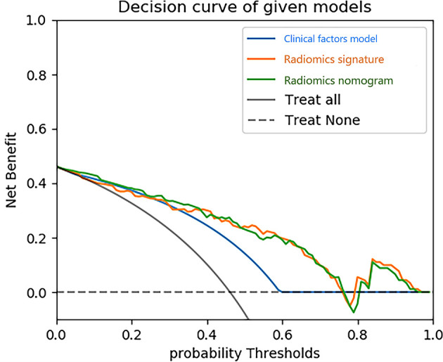
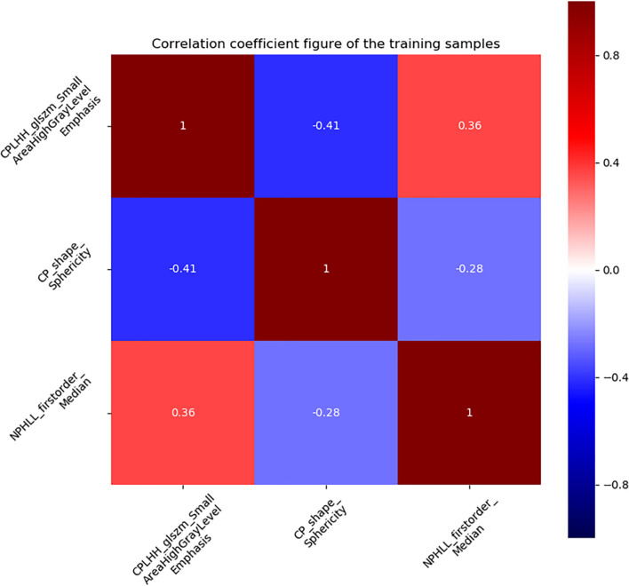
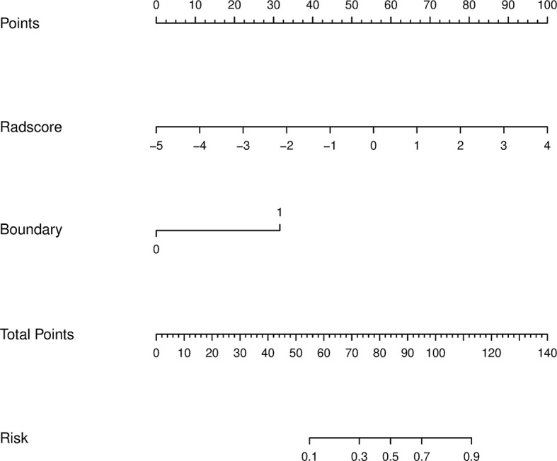
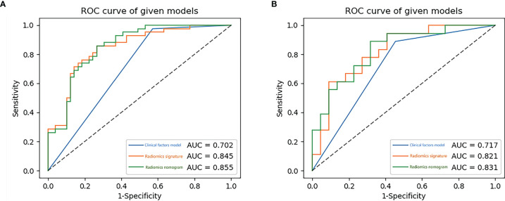
Three validated features were extracted from the CT images and used to construct the radiomics signature. Boundary blurring as an independent risk factor for tumours was used to build clinical factors model. The AUC value of the radiomics nomogram, which was based on the selected clinical factors and Rad-score, were 0.855 and 0.831 in the training and testing sets, respectively. The decision curves of the radiomics nomogram and radiomics signature in the training set indicated an overall net benefit over the clinical factors model.

CONCLUSION

Radiomics nomogram combining clinical factors and radiomics signature is a non-invasive prediction method with a good prediction for pRCC type 1 tumours and type 2 tumours preoperatively and has some significance in guiding clinicians selecting subsequent treatment plans.

摘要

目的

构建基于对比增强CT的影像组学列线图,结合临床因素和影像组学特征,以鉴别1型乳头状肾细胞癌(pRCC)和2型pRCC肿瘤。

方法

共纳入131例患者,其中1型pRCC患者60例,2型pRCC患者71例,并将其分为训练集(n = 91)和测试集(n = 40)。评估患者的人口统计学特征和增强CT影像特征,以建立临床因素模型。通过从皮质髓质期(CMP)和肾实质期(NP)的对比增强CT图像中提取影像组学特征,构建影像组学特征并计算影像组学评分(Rad-score)。然后根据多变量逻辑回归分析,将Rad-score和重要临床因素纳入,构建影像组学列线图。在训练集和测试集上评估临床因素模型、影像组学特征和影像组学列线图的诊断性能。

结果

从CT图像中提取了三个经过验证的特征,并用于构建影像组学特征。将边界模糊作为肿瘤的独立危险因素,用于建立临床因素模型。基于所选临床因素和Rad-score的影像组学列线图在训练集和测试集的AUC值分别为0.855和0.831。训练集中影像组学列线图和影像组学特征的决策曲线表明,其总体净效益优于临床因素模型。

结论

结合临床因素和影像组学特征的影像组学列线图是一种非侵入性预测方法,对术前1型和2型pRCC肿瘤具有良好的预测能力,对指导临床医生选择后续治疗方案具有一定意义。

https://cdn.ncbi.nlm.nih.gov/pmc/blobs/8c17/9204229/497e1521aad1/fonc-12-854979-g009.jpg
https://cdn.ncbi.nlm.nih.gov/pmc/blobs/8c17/9204229/62c9070920d2/fonc-12-854979-g001.jpg
https://cdn.ncbi.nlm.nih.gov/pmc/blobs/8c17/9204229/173a9cb79912/fonc-12-854979-g002.jpg
https://cdn.ncbi.nlm.nih.gov/pmc/blobs/8c17/9204229/7faaf51f8538/fonc-12-854979-g003.jpg
https://cdn.ncbi.nlm.nih.gov/pmc/blobs/8c17/9204229/8eac0617473a/fonc-12-854979-g004.jpg
https://cdn.ncbi.nlm.nih.gov/pmc/blobs/8c17/9204229/8c10266c6a47/fonc-12-854979-g005.jpg
https://cdn.ncbi.nlm.nih.gov/pmc/blobs/8c17/9204229/188dfab5e50c/fonc-12-854979-g006.jpg
https://cdn.ncbi.nlm.nih.gov/pmc/blobs/8c17/9204229/161827fd598d/fonc-12-854979-g007.jpg
https://cdn.ncbi.nlm.nih.gov/pmc/blobs/8c17/9204229/282f60b17efa/fonc-12-854979-g008.jpg
https://cdn.ncbi.nlm.nih.gov/pmc/blobs/8c17/9204229/497e1521aad1/fonc-12-854979-g009.jpg

相似文献

1
Differential Diagnosis of Type 1 and Type 2 Papillary Renal Cell Carcinoma Based on Enhanced CT Radiomics Nomogram.基于增强CT影像组学列线图的1型和2型乳头状肾细胞癌的鉴别诊断
Front Oncol. 2022 Jun 3;12:854979. doi: 10.3389/fonc.2022.854979. eCollection 2022.
2
A Clinical Radiomics Nomogram Was Developed by Integrating Radiomics Signatures and Clinical Variables to Distinguish High-Grade ccRCC from Type 2 pRCC.通过整合影像组学特征和临床变量开发了一种临床影像组学列线图,以区分高级别透明细胞肾细胞癌和2型乳头状肾细胞癌。
J Oncol. 2022 Aug 26;2022:6844349. doi: 10.1155/2022/6844349. eCollection 2022.
3
A CT-based radiomics nomogram for differentiation of small masses (< 4 cm) of renal oncocytoma from clear cell renal cell carcinoma.基于CT的影像组学列线图用于鉴别肾嗜酸细胞瘤与透明细胞肾细胞癌的小肿块(<4cm)。
Abdom Radiol (NY). 2021 Nov;46(11):5240-5249. doi: 10.1007/s00261-021-03213-6. Epub 2021 Jul 15.
4
A CT-based radiomics nomogram for differentiation of renal angiomyolipoma without visible fat from homogeneous clear cell renal cell carcinoma.基于 CT 的影像组学列线图,用于区分无可见脂肪的肾血管平滑肌脂肪瘤与均质透明细胞肾细胞癌。
Eur Radiol. 2020 Feb;30(2):1274-1284. doi: 10.1007/s00330-019-06427-x. Epub 2019 Sep 10.
5
A CT-based radiomics nomogram for differentiation of benign and malignant small renal masses (≤4 cm).基于CT的鉴别肾小肿块(≤4 cm)良恶性的影像组学列线图。
Transl Oncol. 2023 Mar;29:101627. doi: 10.1016/j.tranon.2023.101627. Epub 2023 Jan 31.
6
A CT-based radiomics nomogram for differentiation of renal oncocytoma and chromophobe renal cell carcinoma with a central scar-matched study.基于 CT 的放射组学列线图用于中央瘢痕匹配研究中鉴别肾嗜酸细胞瘤和嫌色细胞肾细胞癌。
Br J Radiol. 2022 Jan 1;95(1129):20210534. doi: 10.1259/bjr.20210534. Epub 2021 Nov 4.
7
A CT-based radiomics nomogram for differentiation of lympho-associated benign and malignant lesions of the parotid gland.基于 CT 的影像组学列线图用于鉴别腮腺淋巴结相关良恶性病变。
Eur Radiol. 2021 May;31(5):2886-2895. doi: 10.1007/s00330-020-07421-4. Epub 2020 Oct 30.
8
A CT-based radiomics nomogram for differentiation of squamous cell carcinoma and non-Hodgkin's lymphoma of the palatine tonsil.基于 CT 的影像组学列线图用于鉴别腭扁桃体的鳞状细胞癌和非霍奇金淋巴瘤。
Eur Radiol. 2022 Jan;32(1):243-253. doi: 10.1007/s00330-021-08153-9. Epub 2021 Jul 8.
9
Multiphase CT radiomics nomogram for preoperatively predicting the WHO/ISUP nuclear grade of small (< 4 cm) clear cell renal cell carcinoma.多相 CT 放射组学列线图,用于术前预测小(<4cm)透明细胞肾细胞癌的 WHO/ISUP 核分级。
BMC Cancer. 2023 Oct 9;23(1):953. doi: 10.1186/s12885-023-11454-5.
10
Extrathyroidal Extension Prediction of Papillary Thyroid Cancer With Computed Tomography Based Radiomics Nomogram: A Multicenter Study.基于计算机断层扫描的影像组学列线图预测甲状腺乳头状癌的甲状腺外侵犯:一项多中心研究
Front Endocrinol (Lausanne). 2022 Jun 1;13:874396. doi: 10.3389/fendo.2022.874396. eCollection 2022.

引用本文的文献

1
A CT-based intratumoral and peritumoral radiomics nomogram for postoperative recurrence risk stratification in localized clear cell renal cell carcinoma.基于CT的瘤内和瘤周影像组学列线图用于局部透明细胞肾细胞癌术后复发风险分层
BMC Med Imaging. 2025 May 16;25(1):167. doi: 10.1186/s12880-025-01715-z.
2
Radiogenomics: a key component of precision cancer medicine.放射组学:精准肿瘤医学的关键组成部分。
Br J Cancer. 2023 Sep;129(5):741-753. doi: 10.1038/s41416-023-02317-8. Epub 2023 Jul 6.

本文引用的文献

1
A CT-based radiomics nomogram for differentiation of renal oncocytoma and chromophobe renal cell carcinoma with a central scar-matched study.基于 CT 的放射组学列线图用于中央瘢痕匹配研究中鉴别肾嗜酸细胞瘤和嫌色细胞肾细胞癌。
Br J Radiol. 2022 Jan 1;95(1129):20210534. doi: 10.1259/bjr.20210534. Epub 2021 Nov 4.
2
Whether histologic subtyping affect the oncological outcomes of patients with papillary renal cell carcinoma: evidence from a systematic review and meta-analysis.组织学亚型是否影响乳头状肾细胞癌患者的肿瘤学结局:一项系统评价和荟萃分析的证据
Transl Androl Urol. 2021 Aug;10(8):3255-3266. doi: 10.21037/tau-21-329.
3
A CT-based radiomics nomogram for differentiation of small masses (< 4 cm) of renal oncocytoma from clear cell renal cell carcinoma.
基于CT的影像组学列线图用于鉴别肾嗜酸细胞瘤与透明细胞肾细胞癌的小肿块(<4cm)。
Abdom Radiol (NY). 2021 Nov;46(11):5240-5249. doi: 10.1007/s00261-021-03213-6. Epub 2021 Jul 15.
4
Radiomics models based on enhanced computed tomography to distinguish clear cell from non-clear cell renal cell carcinomas.基于增强计算机断层扫描的影像组学模型鉴别肾透明细胞癌与非透明细胞癌。
Sci Rep. 2021 Jul 2;11(1):13729. doi: 10.1038/s41598-021-93069-z.
5
Gradient Boosting Decision Tree Algorithm for the Prediction of Postoperative Intraocular Lens Position in Cataract Surgery.用于预测白内障手术中人工晶状体术后位置的梯度提升决策树算法
Transl Vis Sci Technol. 2020 Dec 21;9(13):38. doi: 10.1167/tvst.9.13.38. eCollection 2020 Dec.
6
The effect of the papillary renal cell carcinoma subtype on oncological outcomes.乳头状肾细胞癌亚型对肿瘤学结局的影响。
Sci Rep. 2020 Dec 3;10(1):21073. doi: 10.1038/s41598-020-78174-9.
7
Shape and texture-based radiomics signature on CT effectively discriminates benign from malignant renal masses.基于形状和纹理的 CT 放射组学特征可有效区分良恶性肾肿块。
Eur Radiol. 2021 Feb;31(2):1011-1021. doi: 10.1007/s00330-020-07158-0. Epub 2020 Aug 15.
8
Multiparametric MRI Radiomic Model for Preoperative Predicting WHO/ISUP Nuclear Grade of Clear Cell Renal Cell Carcinoma.用于术前预测透明细胞肾细胞癌WHO/ISUP核级别的多参数MRI影像组学模型
J Magn Reson Imaging. 2020 Nov;52(5):1557-1566. doi: 10.1002/jmri.27182. Epub 2020 May 28.
9
CT texture analysis for the differentiation of papillary renal cell carcinoma subtypes.CT 纹理分析在乳头状肾细胞癌亚型鉴别中的应用。
Abdom Radiol (NY). 2020 Nov;45(11):3860-3868. doi: 10.1007/s00261-020-02588-2.
10
Differentiation of renal cell carcinoma subtypes through MRI-based radiomics analysis.基于 MRI 的影像组学分析对肾细胞癌亚型的鉴别诊断。
Eur Radiol. 2020 Oct;30(10):5738-5747. doi: 10.1007/s00330-020-06896-5. Epub 2020 May 4.