• 文献检索
  • 文档翻译
  • 深度研究
  • 学术资讯
  • Suppr Zotero 插件Zotero 插件
  • 邀请有礼
  • 套餐&价格
  • 历史记录
应用&插件
Suppr Zotero 插件Zotero 插件浏览器插件Mac 客户端Windows 客户端微信小程序
定价
高级版会员购买积分包购买API积分包
服务
文献检索文档翻译深度研究API 文档MCP 服务
关于我们
关于 Suppr公司介绍联系我们用户协议隐私条款
关注我们

Suppr 超能文献

核心技术专利:CN118964589B侵权必究
粤ICP备2023148730 号-1Suppr @ 2026

文献检索

告别复杂PubMed语法,用中文像聊天一样搜索,搜遍4000万医学文献。AI智能推荐,让科研检索更轻松。

立即免费搜索

文件翻译

保留排版,准确专业,支持PDF/Word/PPT等文件格式,支持 12+语言互译。

免费翻译文档

深度研究

AI帮你快速写综述,25分钟生成高质量综述,智能提取关键信息,辅助科研写作。

立即免费体验

一种用于有效分割脑肿瘤的序列机器学习与注意力机制相结合的方法

A Sequential Machine Learning-cum-Attention Mechanism for Effective Segmentation of Brain Tumor.

作者信息

Ali Tahir Mohammad, Nawaz Ali, Ur Rehman Attique, Ahmad Rana Zeeshan, Javed Abdul Rehman, Gadekallu Thippa Reddy, Chen Chin-Ling, Wu Chih-Ming

机构信息

Department of Computer Science, GULF University for Science and Technology, Mishref, Kuwait.

Department of Software Engineering, University of Sialkot, Sialkot, Pakistan.

出版信息

Front Oncol. 2022 Jun 1;12:873268. doi: 10.3389/fonc.2022.873268. eCollection 2022.

DOI:10.3389/fonc.2022.873268
PMID:35719987
原文链接:https://pmc.ncbi.nlm.nih.gov/articles/PMC9202559/
Abstract

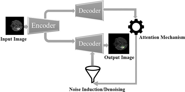
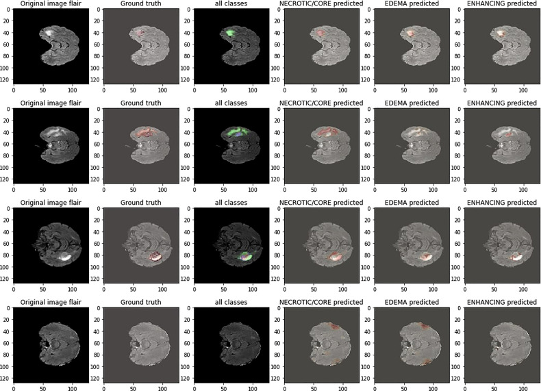
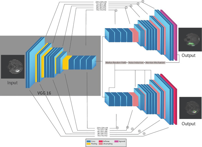
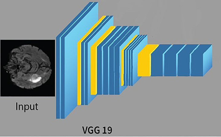
Magnetic resonance imaging is the most generally utilized imaging methodology that permits radiologists to look inside the cerebrum using radio waves and magnets for tumor identification. However, it is tedious and complex to identify the tumorous and nontumorous regions due to the complexity in the tumorous region. Therefore, reliable and automatic segmentation and prediction are necessary for the segmentation of brain tumors. This paper proposes a reliable and efficient neural network variant, i.e., an attention-based convolutional neural network for brain tumor segmentation. Specifically, an encoder part of the UNET is a pre-trained VGG19 network followed by the adjacent decoder parts with an attention gate for segmentation noise induction and a denoising mechanism for avoiding overfitting. The dataset we are using for segmentation is BRATS'20, which comprises four different MRI modalities and one target mask file. The abovementioned algorithm resulted in a dice similarity coefficient of 0.83, 0.86, and 0.90 for enhancing, core, and whole tumors, respectively.

摘要

磁共振成像是最常用的成像方法,它使放射科医生能够利用无线电波和磁体观察大脑内部以识别肿瘤。然而,由于肿瘤区域的复杂性,识别肿瘤和非肿瘤区域既繁琐又复杂。因此,对于脑肿瘤分割来说,可靠且自动的分割和预测是必要的。本文提出了一种可靠且高效的神经网络变体,即用于脑肿瘤分割的基于注意力的卷积神经网络。具体而言,UNET的编码器部分是一个预训练的VGG19网络,其后是相邻的解码器部分,带有用于分割噪声诱导的注意力门和用于避免过拟合的去噪机制。我们用于分割的数据集是BRATS'20,它包含四种不同的MRI模态和一个目标掩码文件。上述算法在增强肿瘤、核心肿瘤和整个肿瘤方面分别产生了0.83、0.86和0.90的骰子相似系数。

https://cdn.ncbi.nlm.nih.gov/pmc/blobs/1d04/9202559/743b620768a2/fonc-12-873268-g005.jpg
https://cdn.ncbi.nlm.nih.gov/pmc/blobs/1d04/9202559/ddd170403323/fonc-12-873268-g001.jpg
https://cdn.ncbi.nlm.nih.gov/pmc/blobs/1d04/9202559/5b31ccadb07c/fonc-12-873268-g002.jpg
https://cdn.ncbi.nlm.nih.gov/pmc/blobs/1d04/9202559/000a19773c5f/fonc-12-873268-g003.jpg
https://cdn.ncbi.nlm.nih.gov/pmc/blobs/1d04/9202559/50010ceb0c4e/fonc-12-873268-g004.jpg
https://cdn.ncbi.nlm.nih.gov/pmc/blobs/1d04/9202559/743b620768a2/fonc-12-873268-g005.jpg

相似文献

1
A Sequential Machine Learning-cum-Attention Mechanism for Effective Segmentation of Brain Tumor.一种用于有效分割脑肿瘤的序列机器学习与注意力机制相结合的方法
Front Oncol. 2022 Jun 1;12:873268. doi: 10.3389/fonc.2022.873268. eCollection 2022.
2
MLRD-Net: 3D multiscale local cross-channel residual denoising network for MRI-based brain tumor segmentation.MLRD-Net:用于基于磁共振成像的脑肿瘤分割的3D多尺度局部跨通道残差去噪网络
Med Biol Eng Comput. 2022 Dec;60(12):3377-3395. doi: 10.1007/s11517-022-02673-2. Epub 2022 Oct 3.
3
A Two-Stage Cascade Model with Variational Autoencoders and Attention Gates for MRI Brain Tumor Segmentation.一种用于MRI脑肿瘤分割的带有变分自编码器和注意力门的两阶段级联模型。
Brainlesion. 2020 Oct;2020:435-447. doi: 10.1007/978-3-030-72084-1_39. Epub 2021 Mar 27.
4
Brain Tumor Segmentation Using Convolutional Neural Networks in MRI Images.基于 MRI 图像的卷积神经网络脑肿瘤分割。
IEEE Trans Med Imaging. 2016 May;35(5):1240-1251. doi: 10.1109/TMI.2016.2538465. Epub 2016 Mar 4.
5
EAR-UNet: A deep learning-based approach for segmentation of tympanic membranes from otoscopic images.基于深度学习的耳镜鼓膜分割方法
Artif Intell Med. 2021 May;115:102065. doi: 10.1016/j.artmed.2021.102065. Epub 2021 Apr 8.
6
Brain tumor segmentation based on deep learning and an attention mechanism using MRI multi-modalities brain images.基于深度学习和注意力机制的 MRI 多模态脑图像脑肿瘤分割。
Sci Rep. 2021 May 25;11(1):10930. doi: 10.1038/s41598-021-90428-8.
7
An Efficient Implementation of Deep Convolutional Neural Networks for MRI Segmentation.一种用于 MRI 分割的高效卷积神经网络实现。
J Digit Imaging. 2018 Oct;31(5):738-747. doi: 10.1007/s10278-018-0062-2.
8
Efficient U-Net Architecture with Multiple Encoders and Attention Mechanism Decoders for Brain Tumor Segmentation.用于脑肿瘤分割的具有多个编码器和注意力机制解码器的高效U-Net架构
Diagnostics (Basel). 2023 Feb 24;13(5):872. doi: 10.3390/diagnostics13050872.
9
MM-UNet: A multimodality brain tumor segmentation network in MRI images.MM-UNet:一种用于磁共振成像(MRI)图像的多模态脑肿瘤分割网络。
Front Oncol. 2022 Aug 18;12:950706. doi: 10.3389/fonc.2022.950706. eCollection 2022.
10
An Intelligent Diagnosis Method of Brain MRI Tumor Segmentation Using Deep Convolutional Neural Network and SVM Algorithm.基于深度卷积神经网络和支持向量机算法的脑 MRI 肿瘤分割智能诊断方法。
Comput Math Methods Med. 2020 Jul 14;2020:6789306. doi: 10.1155/2020/6789306. eCollection 2020.

引用本文的文献

1
G-Net: Implementing an enhanced brain tumor segmentation framework using semantic segmentation design.G-Net:使用语义分割设计实现增强型脑肿瘤分割框架。
PLoS One. 2024 Aug 6;19(8):e0308236. doi: 10.1371/journal.pone.0308236. eCollection 2024.
2
Enhancing brain tumor segmentation in MRI images using the IC-net algorithm framework.利用 IC-net 算法框架增强 MRI 图像中的脑肿瘤分割。
Sci Rep. 2024 Jul 8;14(1):15660. doi: 10.1038/s41598-024-66314-4.
3
Enhancing cervical cancer detection and robust classification through a fusion of deep learning models.

本文引用的文献

1
FL-PMI: Federated Learning-Based Person Movement Identification through Wearable Devices in Smart Healthcare Systems.基于联邦学习的智能医疗系统中可穿戴设备的人员移动识别
Sensors (Basel). 2022 Feb 11;22(4):1377. doi: 10.3390/s22041377.
2
Missing data imputation on biomedical data using deeply learned clustering and L2 regularized regression based on symmetric uncertainty.基于对称不确定性,使用深度聚类和L2正则化回归对生物医学数据进行缺失数据插补。
Artif Intell Med. 2022 Jan;123:102214. doi: 10.1016/j.artmed.2021.102214. Epub 2021 Dec 6.
3
A deep learning method for automatic segmentation of the bony orbit in MRI and CT images.
通过深度学习模型融合提高宫颈癌检测和稳健分类。
Sci Rep. 2024 May 11;14(1):10812. doi: 10.1038/s41598-024-61063-w.
4
Privacy Preserved Cervical Cancer Detection Using Convolutional Neural Networks Applied to Pap Smear Images.基于卷积神经网络的巴氏涂片图像隐私保护宫颈癌检测
Comput Math Methods Med. 2023 Jul 8;2023:9676206. doi: 10.1155/2023/9676206. eCollection 2023.
5
Identifying Thematics in a Brain-Computer Interface Research.识别脑机接口研究中的主题。
Comput Intell Neurosci. 2023 Jan 4;2023:2793211. doi: 10.1155/2023/2793211. eCollection 2023.
6
Application of Machine Learning in Intelligent Medical Image Diagnosis and Construction of Intelligent Service Process.机器学习在智能医学影像诊断中的应用及智能服务流程的构建。
Comput Intell Neurosci. 2022 Dec 28;2022:9152605. doi: 10.1155/2022/9152605. eCollection 2022.
7
An efficient computer vision-based approach for acute lymphoblastic leukemia prediction.一种基于计算机视觉的急性淋巴细胞白血病高效预测方法。
Front Comput Neurosci. 2022 Nov 24;16:1083649. doi: 10.3389/fncom.2022.1083649. eCollection 2022.
8
Auxiliary Segmentation Method of Osteosarcoma MRI Image Based on Transformer and U-Net.基于 Transformer 和 U-Net 的骨肉瘤 MRI 图像辅助分割方法。
Comput Intell Neurosci. 2022 Nov 14;2022:9990092. doi: 10.1155/2022/9990092. eCollection 2022.
9
Ensemble deep learning for brain tumor detection.用于脑肿瘤检测的集成深度学习
Front Comput Neurosci. 2022 Sep 2;16:1005617. doi: 10.3389/fncom.2022.1005617. eCollection 2022.
10
Segmentation and Classification of Encephalon Tumor by Applying Improved Fast and Robust FCM Algorithm with PSO-Based ELM Technique.应用基于 PSO 的 ELM 技术改进的快速鲁棒 FCM 算法对脑肿瘤进行分割和分类。
Comput Intell Neurosci. 2022 Jul 31;2022:2664901. doi: 10.1155/2022/2664901. eCollection 2022.
一种用于 MRI 和 CT 图像中骨眼眶自动分割的深度学习方法。
Sci Rep. 2021 Jul 1;11(1):13693. doi: 10.1038/s41598-021-93227-3.
4
Improved U-Net architecture with VGG-16 for brain tumor segmentation.基于 VGG-16 的改进 U-Net 架构用于脑肿瘤分割。
Phys Eng Sci Med. 2021 Sep;44(3):703-712. doi: 10.1007/s13246-021-01019-w. Epub 2021 May 28.
5
Context aware deep learning for brain tumor segmentation, subtype classification, and survival prediction using radiology images.基于放射影像的脑肿瘤分割、亚型分类和生存预测的上下文感知深度学习
Sci Rep. 2020 Nov 12;10(1):19726. doi: 10.1038/s41598-020-74419-9.
6
High tissue contrast image synthesis via multistage attention-GAN: Application to segmenting brain MR scans.通过多阶段注意生成对抗网络进行高组织对比图像合成:在脑磁共振扫描分割中的应用。
Neural Netw. 2020 Dec;132:43-52. doi: 10.1016/j.neunet.2020.08.014. Epub 2020 Aug 18.
7
DeepSeg: deep neural network framework for automatic brain tumor segmentation using magnetic resonance FLAIR images.DeepSeg:基于磁共振 FLAIR 图像的自动脑肿瘤分割的深度神经网络框架。
Int J Comput Assist Radiol Surg. 2020 Jun;15(6):909-920. doi: 10.1007/s11548-020-02186-z. Epub 2020 May 5.
8
BrainMRNet: Brain tumor detection using magnetic resonance images with a novel convolutional neural network model.BrainMRNet:使用新型卷积神经网络模型对磁共振图像进行脑肿瘤检测。
Med Hypotheses. 2020 Jan;134:109531. doi: 10.1016/j.mehy.2019.109531. Epub 2019 Dec 17.
9
Brain Tumor Segmentation and Survival Prediction Using Multimodal MRI Scans With Deep Learning.使用深度学习的多模态磁共振成像扫描进行脑肿瘤分割与生存预测
Front Neurosci. 2019 Aug 16;13:810. doi: 10.3389/fnins.2019.00810. eCollection 2019.
10
A hybrid of whale optimization and late acceptance hill climbing based imputation to enhance classification performance in electronic health records.基于鲸鱼优化算法和后期接受爬山算法的混合插补方法提高电子健康记录中的分类性能。
J Biomed Inform. 2019 Jun;94:103190. doi: 10.1016/j.jbi.2019.103190. Epub 2019 May 2.