Whitaker Emmett E, Johnson Abbie C, Tremble Sarah M, McGinn Conor, DeLance Nicole, Cipolla Marilyn J
Department of Anesthesiology, University of Vermont Larner College of Medicine, Burlington, VT, United States.
Department of Neurological Sciences, University of Vermont Larner College of Medicine, Burlington, VT, United States.
Front Physiol. 2022 Jun 6;13:924908. doi: 10.3389/fphys.2022.924908. eCollection 2022.
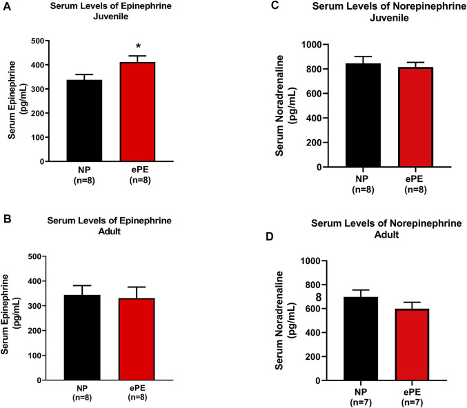
Preeclampsia is a hypertensive disorder of pregnancy that causes significant, long term cardiovascular effects for both the mother and offspring. A previous study demonstrated that middle cerebral arteries in offspring from an experimental rat model of preeclampsia were smaller, stiffer, and did not enlarge over the course of maturation, suggesting potential hemodynamic alterations in these offspring. Here we investigated the effect of experimental preeclampsia on cerebral blood flow autoregulation in juvenile and adult offspring that were born from normal pregnant or experimentally preeclamptic rats. Relative cerebral blood flow was measured using laser Doppler flowmetry, and cerebral blood flow autoregulation curves were constructed by raising blood pressure and controlled hemorrhage to lower blood pressure. Immunohistochemistry was used to assess middle cerebral artery size. Heart rate and blood pressure were measured in awake adult offspring using implanted radiotelemetry. Serum epinephrine was measured using enzyme-linked immunosorbent assay. Offspring from both groups showed maturation of cerebral blood flow autoregulation as offspring aged from juvenile to adulthood as demonstrated by the wider autoregulatory plateau. Experimental preeclampsia did not affect cerebral blood flow autoregulation in juvenile offspring, and it had no effect on cerebral blood flow autoregulation in adult offspring over the lower range of blood pressures. However, experimental preeclampsia caused a right shift in the upper range of blood pressures in adult offspring (compared to normal pregnant). Structurally, middle cerebral arteries from normal pregnant offspring demonstrated growth with aging, while middle cerebral arteries from experimentally preeclamptic offspring did not, and by adulthood normal pregnant offspring had significantly larger middle cerebral arteries. Middle cerebral artery lumen diameters did not significantly change as offspring aged. Serum epinephrine was elevated in juvenile experimentally preeclamptic offspring, and a greater degree of hemorrhage was required to induce hypotension, suggesting increased sympathetic activity. Finally, despite no evidence of increased sympathetic activity, adult experimentally preeclamptic offspring were found to have persistently higher heart rate. These results demonstrate a significant effect of experimental preeclampsia on the upper range of autoregulation and cerebrovascular structure in juvenile and adult offspring that could have an important influence on brain perfusion under conditions of hypo and/or hypertension.
子痫前期是一种妊娠期高血压疾病,会对母亲和后代造成重大的长期心血管影响。先前的一项研究表明,子痫前期实验大鼠模型后代的大脑中动脉较小、较硬,且在成熟过程中不会增大,这表明这些后代可能存在血流动力学改变。在此,我们研究了实验性子痫前期对正常妊娠或实验性子痫前期大鼠所产后代幼年和成年期脑血流自动调节的影响。使用激光多普勒血流仪测量相对脑血流量,并通过升高血压和控制性出血降低血压来构建脑血流自动调节曲线。采用免疫组织化学法评估大脑中动脉大小。使用植入式无线电遥测技术测量清醒成年后代的心率和血压。采用酶联免疫吸附测定法测量血清肾上腺素。两组后代随着从幼年到成年的年龄增长,脑血流自动调节均表现出成熟,表现为自动调节平台变宽。实验性子痫前期对幼年后代的脑血流自动调节没有影响,在较低血压范围内对成年后代的脑血流自动调节也没有影响。然而,实验性子痫前期导致成年后代血压上限右移(与正常妊娠相比)。在结构上,正常妊娠后代的大脑中动脉随年龄增长而生长,而实验性子痫前期后代的大脑中动脉则不然,到成年时,正常妊娠后代的大脑中动脉明显更大。大脑中动脉管腔直径并未随着后代年龄增长而显著变化。实验性子痫前期幼年后代血清肾上腺素升高,诱导低血压需要更大程度的出血,提示交感神经活动增加。最后,尽管没有证据表明交感神经活动增加,但发现实验性子痫前期成年后代的心率持续较高。这些结果表明,实验性子痫前期对幼年和成年后代的自动调节上限和脑血管结构有显著影响,这可能对低血压和/或高血压情况下的脑灌注产生重要影响。