Su Hao, Kwok Ka-Wai, Cleary Kevin, Iordachita Iulian, Cavusoglu M Cenk, Desai Jaydev P, Fischer Gregory S
Department of Mechanical and Aerospace Engineering, North Carolina State University, Raleigh, NC 27695 USA.
Department of Mechanical Engineering, The University of Hong Kong, Hong Kong.
Proc IEEE Inst Electr Electron Eng. 2022 Jul;110(7):968-992. doi: 10.1109/jproc.2022.3169146. Epub 2022 May 3.
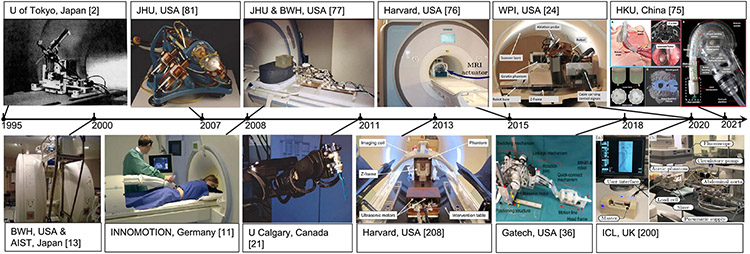
Magnetic resonance imaging (MRI) can provide high-quality 3-D visualization of target anatomy, surrounding tissue, and instrumentation, but there are significant challenges in harnessing it for effectively guiding interventional procedures. Challenges include the strong static magnetic field, rapidly switching magnetic field gradients, high-power radio frequency pulses, sensitivity to electrical noise, and constrained space to operate within the bore of the scanner. MRI has a number of advantages over other medical imaging modalities, including no ionizing radiation, excellent soft-tissue contrast that allows for visualization of tumors and other features that are not readily visible by other modalities, true 3-D imaging capabilities, including the ability to image arbitrary scan plane geometry or perform volumetric imaging, and capability for multimodality sensing, including diffusion, dynamic contrast, blood flow, blood oxygenation, temperature, and tracking of biomarkers. The use of robotic assistants within the MRI bore, alongside the patient during imaging, enables intraoperative MR imaging (iMRI) to guide a surgical intervention in a closed-loop fashion that can include tracking of tissue deformation and target motion, localization of instrumentation, and monitoring of therapy delivery. With the ever-expanding clinical use of MRI, MRI-compatible robotic systems have been heralded as a new approach to assist interventional procedures to allow physicians to treat patients more accurately and effectively. Deploying robotic systems inside the bore synergizes the visual capability of MRI and the manipulation capability of robotic assistance, resulting in a closed-loop surgery architecture. This article details the challenges and history of robotic systems intended to operate in an MRI environment and outlines promising clinical applications and associated state-of-the-art MRI-compatible robotic systems and technology for making this possible.
磁共振成像(MRI)能够提供高质量的目标解剖结构、周围组织及器械的三维可视化图像,但在将其有效用于引导介入手术方面存在重大挑战。这些挑战包括强静磁场、快速切换的磁场梯度、高功率射频脉冲、对电噪声的敏感性以及在扫描孔内操作空间受限等。与其他医学成像模态相比,MRI具有诸多优势,包括无电离辐射、软组织对比度极佳,能够显示肿瘤及其他用其他模态不易看到的特征、具备真正的三维成像能力,包括能够对任意扫描平面几何形状进行成像或进行容积成像,以及具备多模态传感能力,包括扩散、动态对比、血流、血氧、温度和生物标志物追踪等。在MRI扫描孔内使用机器人助手,在成像过程中与患者一起,可使术中磁共振成像(iMRI)以闭环方式引导手术干预,这可以包括组织变形和目标运动追踪、器械定位以及治疗输送监测。随着MRI在临床上的应用不断扩展,与MRI兼容的机器人系统被誉为一种辅助介入手术的新方法,使医生能够更准确、有效地治疗患者。在扫描孔内部署机器人系统可使MRI的视觉能力与机器人辅助的操作能力协同作用,从而形成一种闭环手术架构。本文详细介绍了旨在在MRI环境中运行的机器人系统所面临的挑战和发展历程,并概述了有前景的临床应用以及相关的最先进的与MRI兼容的机器人系统和技术,以使这一切成为可能。