Department of Radiology, Valenciennes Hospital Center, 114 Avenue Desandrouin, 59300, Valenciennes, France.
Department of Public Health, EA 2694, Lille University, 1 Place de Verdun, 59045, Lille Cedex, France.
Breast Cancer. 2022 Nov;29(6):967-977. doi: 10.1007/s12282-022-01375-9. Epub 2022 Jun 28.
To demonstrate that radiologists, with the help of artificial intelligence (AI), are able to better classify screening mammograms into the correct breast imaging reporting and data system (BI-RADS) category, and as a secondary objective, to explore the impact of AI on cancer detection and mammogram interpretation time.
A multi-reader, multi-case study with cross-over design, was performed, including 314 mammograms. Twelve radiologists interpreted the examinations in two sessions delayed by a 4 weeks wash-out period with and without AI support. For each breast of each mammogram, they had to mark the most suspicious lesion (if any) and assign it with a forced BI-RADS category and a level of suspicion or "continuous BI-RADS 100". Cohen's kappa correlation coefficient evaluating the inter-observer agreement for BI-RADS category per breast, and the area under the receiver operating characteristic curve (AUC), were used as metrics and analyzed.
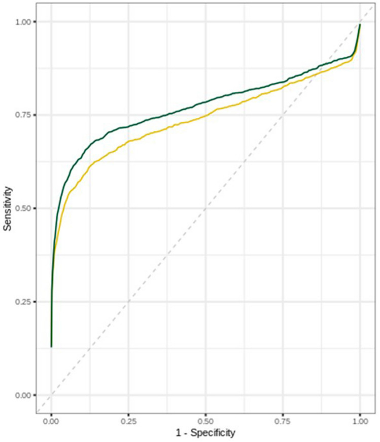
On average, the quadratic kappa coefficient increased significantly when using AI for all readers [κ = 0.549, 95% CI (0.528-0.571) without AI and κ = 0.626, 95% CI (0.607-0.6455) with AI]. AUC was significantly improved when using AI (0.74 vs 0.77, p = 0.004). Reading time was not significantly affected for all readers (106 s without AI and vs 102 s with AI; p = 0.754).
When using AI, radiologists were able to better assign mammograms with the correct BI-RADS category without slowing down the interpretation time.
证明放射科医生在人工智能(AI)的帮助下,能够更好地将筛查性乳房 X 光照片分类到正确的乳房影像报告和数据系统(BI-RADS)类别中;并作为次要目标,探索 AI 对癌症检测和乳房 X 光照片解读时间的影响。
采用多读者、多病例交叉设计进行了一项研究,纳入了 314 例乳房 X 光照片。12 名放射科医生在相隔 4 周的洗脱期内,分两次对这些检查进行解读,一次有 AI 支持,一次没有。对于每张乳房 X 光照片的每只乳房,他们都必须标记出最可疑的病变(如果有),并为其分配强制 BI-RADS 类别和可疑程度(或“连续 BI-RADS 100”)。采用 Cohen 氏 κ 相关系数评估每只乳房的 BI-RADS 类别观察者间一致性,并分析其曲线下面积(AUC)。
平均而言,当所有读者都使用 AI 时,二次 κ 值显著增加[κ=0.549,95%可信区间(0.528-0.571)无 AI 时,κ=0.626,95%可信区间(0.607-0.6455)有 AI 时]。使用 AI 时 AUC 显著提高(0.74 比 0.77,p=0.004)。所有读者的阅读时间均无显著变化(无 AI 时为 106 秒,有 AI 时为 102 秒;p=0.754)。
使用 AI 时,放射科医生能够更好地为乳房 X 光照片分配正确的 BI-RADS 类别,而不会减缓解读时间。