• 文献检索
  • 文档翻译
  • 深度研究
  • 学术资讯
  • Suppr Zotero 插件Zotero 插件
  • 邀请有礼
  • 套餐&价格
  • 历史记录
应用&插件
Suppr Zotero 插件Zotero 插件浏览器插件Mac 客户端Windows 客户端微信小程序
定价
高级版会员购买积分包购买API积分包
服务
文献检索文档翻译深度研究API 文档MCP 服务
关于我们
关于 Suppr公司介绍联系我们用户协议隐私条款
关注我们

Suppr 超能文献

核心技术专利:CN118964589B侵权必究
粤ICP备2023148730 号-1Suppr @ 2026

文献检索

告别复杂PubMed语法,用中文像聊天一样搜索,搜遍4000万医学文献。AI智能推荐,让科研检索更轻松。

立即免费搜索

文件翻译

保留排版,准确专业,支持PDF/Word/PPT等文件格式,支持 12+语言互译。

免费翻译文档

深度研究

AI帮你快速写综述,25分钟生成高质量综述,智能提取关键信息,辅助科研写作。

立即免费体验

使用正电子发射断层扫描(PET)成像技术在头颈癌中研究缺氧与肿瘤体积的关系——一项范围综述

Hypoxia in relationship to tumor volume using hypoxia PET-imaging in head & neck cancer - A scoping review.

作者信息

Hildingsson Sofia, Gebre-Medhin Maria, Zschaeck Sebastian, Adrian Gabriel

机构信息

Division of Oncology and Pathology, Clinical Sciences, Lund University, Lund, Sweden.

Department of Hematology, Oncology and Radiation Physics, Skåne University Hospital, Lund University, Lund, Sweden.

出版信息

Clin Transl Radiat Oncol. 2022 Jun 15;36:40-46. doi: 10.1016/j.ctro.2022.06.004. eCollection 2022 Sep.

DOI:10.1016/j.ctro.2022.06.004
PMID:35769424
原文链接:https://pmc.ncbi.nlm.nih.gov/articles/PMC9234341/
Abstract

BACKGROUND

Hypoxia and large tumor volumes are negative prognostic factors for patients with head and neck squamous cell carcinoma (HNSCC) treated with radiation therapy (RT). PET-scanning with specific hypoxia-tracers (hypoxia-PET) can be used to non-invasively assess hypoxic tumor volume. Primary tumor volume is readily available for patients undergoing RT. However, the relationship between hypoxic volume and primary tumor volume is yet an open question. The current study investigates the hypotheses that larger tumors contain both a larger hypoxic volume and a higher hypoxic fraction.

METHODS

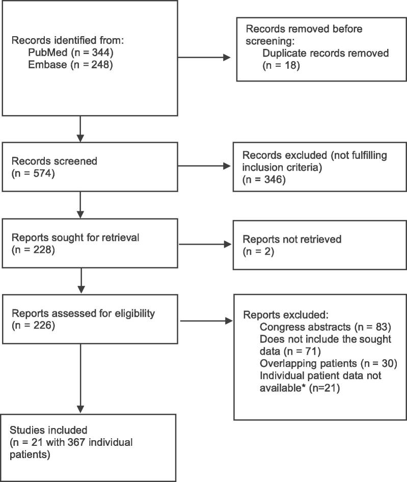
PubMed and Embase were systematically searched to identify articles fulfilling the predefined criteria. Individual tumor data (primary tumor volume and hypoxic volume/fraction) was extracted. Relationship between hypoxic volume and primary tumor volume was investigated by linear regression. The correlation between hypoxic fraction and log(primary tumor volume) was determined for each cohort and in a pooled analysis individual regression slopes and coefficients of determination (R) were weighted according to cohort size.

RESULTS

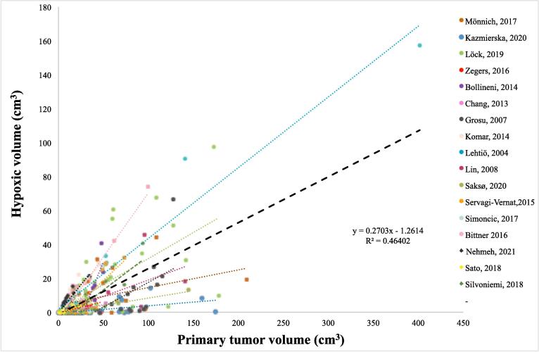
21 relevant articles were identified and individual data from 367 patients was extracted, out of which 323 patients from 17 studies had quantifiable volumes of interest. A correlation between primary tumor volume and PET-determined hypoxic volume was found ( Larger tumors had a significantly higher fraction of hypoxia compared with smaller tumors (.01). The weighted analysis of all studies revealed that for each doubling of the tumor volume, the hypoxic fraction increased by four percentage points.

CONCLUSION

This study shows correlations between primary tumor volume and hypoxic volume as well as primary tumor volume and the hypoxic fraction in patients with HNSCC. The findings suggest that not only do large tumors contain more cancer cells, they also have a higher proportion of potentially radioresistant hypoxic cells. This knowledge can be important when individualizing RT.

摘要

背景

缺氧和大肿瘤体积是接受放射治疗(RT)的头颈部鳞状细胞癌(HNSCC)患者的不良预后因素。使用特定缺氧示踪剂的PET扫描(缺氧PET)可用于无创评估缺氧肿瘤体积。接受RT的患者的原发肿瘤体积很容易获得。然而,缺氧体积与原发肿瘤体积之间的关系仍是一个悬而未决的问题。当前研究调查了以下假设:更大的肿瘤既包含更大的缺氧体积,也包含更高的缺氧分数。

方法

系统检索PubMed和Embase以识别符合预定义标准的文章。提取个体肿瘤数据(原发肿瘤体积和缺氧体积/分数)。通过线性回归研究缺氧体积与原发肿瘤体积之间的关系。确定每个队列中缺氧分数与log(原发肿瘤体积)之间的相关性,并在汇总分析中根据队列大小对个体回归斜率和决定系数(R)进行加权。

结果

识别出21篇相关文章,并提取了367例患者的个体数据,其中来自17项研究的323例患者有可量化的感兴趣体积。发现原发肿瘤体积与PET确定的缺氧体积之间存在相关性(与较小肿瘤相比,较大肿瘤的缺氧分数显著更高(.01)。所有研究的加权分析显示,肿瘤体积每增加一倍,缺氧分数增加4个百分点。

结论

本研究显示了HNSCC患者原发肿瘤体积与缺氧体积以及原发肿瘤体积与缺氧分数之间的相关性。研究结果表明,大肿瘤不仅包含更多癌细胞,还具有更高比例的潜在放射抗性缺氧细胞。在放疗个体化时,这一知识可能很重要。

https://cdn.ncbi.nlm.nih.gov/pmc/blobs/2d62/9234341/94bf917a8bc0/gr2a.jpg
https://cdn.ncbi.nlm.nih.gov/pmc/blobs/2d62/9234341/632d7549b51d/gr1.jpg
https://cdn.ncbi.nlm.nih.gov/pmc/blobs/2d62/9234341/94bf917a8bc0/gr2a.jpg
https://cdn.ncbi.nlm.nih.gov/pmc/blobs/2d62/9234341/632d7549b51d/gr1.jpg
https://cdn.ncbi.nlm.nih.gov/pmc/blobs/2d62/9234341/94bf917a8bc0/gr2a.jpg

相似文献

1
Hypoxia in relationship to tumor volume using hypoxia PET-imaging in head & neck cancer - A scoping review.使用正电子发射断层扫描(PET)成像技术在头颈癌中研究缺氧与肿瘤体积的关系——一项范围综述
Clin Transl Radiat Oncol. 2022 Jun 15;36:40-46. doi: 10.1016/j.ctro.2022.06.004. eCollection 2022 Sep.
2
Influence of FAZA PET hypoxia and HPV-status for the outcome of head and neck squamous cell carcinoma (HNSCC) treated with radiotherapy: Long-term results from the DAHANCA 24 trial (NCT01017224).FAZA PET 乏氧及 HPV 状态对接受放疗的头颈部鳞癌(HNSCC)患者预后的影响:来自 DAHANCA 24 试验的长期结果(NCT01017224)。
Radiother Oncol. 2020 Oct;151:126-133. doi: 10.1016/j.radonc.2020.08.006. Epub 2020 Aug 14.
3
A prospective clinical study of ¹⁸F-FAZA PET-CT hypoxia imaging in head and neck squamous cell carcinoma before and during radiation therapy.¹⁸F-FAZA PET-CT 乏氧成像在头颈部鳞状细胞癌放疗前及放疗期间的前瞻性临床研究。
Eur J Nucl Med Mol Imaging. 2014 Aug;41(8):1544-52. doi: 10.1007/s00259-014-2730-x. Epub 2014 Feb 26.
4
The utility of multiparametric MRI to characterize hypoxic tumor subvolumes in comparison to FMISO PET/CT. Consequences for diagnosis and chemoradiation treatment planning in head and neck cancer.与氟代吗啉硝唑正电子发射断层扫描/计算机断层扫描(FMISO PET/CT)相比,多参数磁共振成像(MRI)对缺氧肿瘤亚体积进行特征描述的效用。对头颈部癌诊断和放化疗治疗计划的影响。
Radiother Oncol. 2020 Sep;150:128-135. doi: 10.1016/j.radonc.2020.06.013. Epub 2020 Jun 13.
5
A novel approach to overcome hypoxic tumor resistance: Cu-ATSM-guided intensity-modulated radiation therapy.一种克服缺氧肿瘤抗性的新方法:铜-ATSM引导的调强放射治疗。
Int J Radiat Oncol Biol Phys. 2001 Mar 15;49(4):1171-82. doi: 10.1016/s0360-3016(00)01433-4.
6
Residual tumour hypoxia in head-and-neck cancer patients undergoing primary radiochemotherapy, final results of a prospective trial on repeat FMISO-PET imaging.接受原发性放化疗的头颈癌患者的残余肿瘤缺氧情况:一项关于重复氟代脱氧吗啉硝唑正电子发射断层扫描成像的前瞻性试验的最终结果
Radiother Oncol. 2017 Sep;124(3):533-540. doi: 10.1016/j.radonc.2017.08.010. Epub 2017 Aug 23.
7
Overlap of highly FDG-avid and FMISO hypoxic tumor subvolumes in patients with head and neck cancer.头颈部癌患者中高氟代脱氧葡萄糖摄取(FDG-avid)和氟米索(FMISO)低氧肿瘤亚体积的重叠情况。
Acta Oncol. 2017 Nov;56(11):1577-1582. doi: 10.1080/0284186X.2017.1363910. Epub 2017 Aug 29.
8
Evaluation of quantitative imaging parameters in head and neck squamous cell carcinoma.头颈部鳞状细胞癌定量成像参数的评估。
Q J Nucl Med Mol Imaging. 2022 Jun;66(2):162-170. doi: 10.23736/S1824-4785.19.03179-0. Epub 2019 Sep 5.
9
PET measured hypoxia and MRI parameters in re-irradiated head and neck squamous cell carcinomas: findings of a prospective pilot study.PET 测量复发性头颈部鳞状细胞癌的缺氧和 MRI 参数:一项前瞻性试点研究的结果。
F1000Res. 2020 Nov 19;9:1350. doi: 10.12688/f1000research.27303.2. eCollection 2020.
10
PET-based quantification of statistical properties of hypoxic tumor subvolumes in head and neck cancer.基于正电子发射断层扫描(PET)对头颈部癌缺氧肿瘤子体积统计特性的量化分析
Phys Med. 2016 Jan;32(1):23-35. doi: 10.1016/j.ejmp.2015.12.006. Epub 2015 Dec 31.

引用本文的文献

1
Imaging hypoxia for head and neck cancer: current status, challenges, and prospects.头颈部癌的缺氧成像:现状、挑战与前景
Theranostics. 2025 Jul 11;15(16):8012-8030. doi: 10.7150/thno.112781. eCollection 2025.
2
FAP-Directed Imaging and Therapy in Head and Neck Cancer of Unknown Primary.未知原发灶头颈部癌的FAP导向成像与治疗
Cancers (Basel). 2025 Jun 30;17(13):2205. doi: 10.3390/cancers17132205.
3
Cyanine dyes in the mitochondria-targeting photodynamic and photothermal therapy.用于线粒体靶向光动力和光热疗法的菁染料

本文引用的文献

1
Primary tumor volume and prognosis for patients with p16-positive and p16-negative oropharyngeal squamous cell carcinoma treated with radiation therapy.放疗治疗 p16 阳性和 p16 阴性口咽鳞癌患者的原发肿瘤体积与预后。
Radiat Oncol. 2022 Jun 14;17(1):107. doi: 10.1186/s13014-022-02074-7.
2
Comparison of FDG and FMISO uptakes and distributions in head and neck squamous cell cancer tumors.头颈部鳞状细胞癌肿瘤中氟代脱氧葡萄糖(FDG)与氟代吗啉代硝唑(FMISO)摄取及分布的比较
EJNMMI Res. 2021 Apr 14;11(1):38. doi: 10.1186/s13550-021-00767-w.
3
Precision Radiotherapy: Reduction in Radiation for Oropharyngeal Cancer in the 30 ROC Trial.
Commun Chem. 2024 Aug 13;7(1):180. doi: 10.1038/s42004-024-01256-6.
4
Diffuse reflectance spectroscopy for optical characterizations of orthotopic head and neck cancer models .用于原位头颈癌模型光学表征的漫反射光谱法
Biomed Opt Express. 2024 Jun 10;15(7):4176-4189. doi: 10.1364/BOE.528608. eCollection 2024 Jul 1.
5
E-Cadherin Induces Serine Synthesis to Support Progression and Metastasis of Breast Cancer.E-钙黏蛋白诱导丝氨酸合成以支持乳腺癌的进展和转移。
Cancer Res. 2024 Sep 4;84(17):2820-2835. doi: 10.1158/0008-5472.CAN-23-3082.
6
Implementation of Oxygen Enhanced Magnetic Resonance Imaging (OE-MRI) and a Pilot Genomic Study of Hypoxia in Bladder Cancer Xenografts.氧增强磁共振成像(OE-MRI)的实现及膀胱癌异种移植模型中缺氧的初步基因组学研究。
Cancer Genomics Proteomics. 2024 Jul-Aug;21(4):380-387. doi: 10.21873/cgp.20455.
7
Modulating cancer mechanopathology to restore vascular function and enhance immunotherapy.调节癌症机制病理学以恢复血管功能并增强免疫疗法。
Cell Rep Med. 2024 Jul 16;5(7):101626. doi: 10.1016/j.xcrm.2024.101626. Epub 2024 Jun 28.
8
Advancing cancer drug development with mechanistic mathematical modeling: bridging the gap between theory and practice.运用机制数学建模推进癌症药物研发:弥合理论与实践之间的差距。
J Pharmacokinet Pharmacodyn. 2024 Dec;51(6):581-604. doi: 10.1007/s10928-024-09930-x. Epub 2024 Jun 21.
9
Rethinking the potential role of dose painting in personalized ultra-fractionated stereotactic adaptive radiotherapy.重新思考剂量勾画在个体化超分割立体定向自适应放射治疗中的潜在作用。
Front Oncol. 2024 Mar 20;14:1357790. doi: 10.3389/fonc.2024.1357790. eCollection 2024.
10
Bioengineering of a tumour-stroma 3D-tumouroid co-culture model of hypopharyngeal cancer.构建下咽癌肿瘤基质三维类器官共培养模型的生物工程技术。
Biol Open. 2023 May 15;12(5). doi: 10.1242/bio.059949. Epub 2023 May 17.
精准放疗:30 个 ROC 试验中减少口咽癌的放射剂量。
J Natl Cancer Inst. 2021 Jun 1;113(6):742-751. doi: 10.1093/jnci/djaa184.
4
Squamous cell carcinoma of the oral cavity, larynx, oropharynx and hypopharynx: EHNS-ESMO-ESTRO Clinical Practice Guidelines for diagnosis, treatment and follow-up.口腔、喉、口咽和下咽鳞状细胞癌:EHNS-ESMO-ESTRO诊断、治疗及随访临床实践指南
Ann Oncol. 2020 Nov;31(11):1462-1475. doi: 10.1016/j.annonc.2020.07.011. Epub 2020 Oct 23.
5
Influence of FAZA PET hypoxia and HPV-status for the outcome of head and neck squamous cell carcinoma (HNSCC) treated with radiotherapy: Long-term results from the DAHANCA 24 trial (NCT01017224).FAZA PET 乏氧及 HPV 状态对接受放疗的头颈部鳞癌(HNSCC)患者预后的影响:来自 DAHANCA 24 试验的长期结果(NCT01017224)。
Radiother Oncol. 2020 Oct;151:126-133. doi: 10.1016/j.radonc.2020.08.006. Epub 2020 Aug 14.
6
Individual patient data meta-analysis of FMISO and FAZA hypoxia PET scans from head and neck cancer patients undergoing definitive radio-chemotherapy.头颈部癌症患者行根治性放化疗时 FMISO 和 FAZA 乏氧 PET 扫描的个体患者数据分析的荟萃分析。
Radiother Oncol. 2020 Aug;149:189-196. doi: 10.1016/j.radonc.2020.05.022. Epub 2020 May 15.
7
AKT inhibition as a strategy for targeting hypoxic HPV-positive HNSCC.AKT 抑制作为一种针对低氧 HPV 阳性头颈部鳞状细胞癌的靶向策略。
Radiother Oncol. 2020 Aug;149:1-7. doi: 10.1016/j.radonc.2020.04.048. Epub 2020 Apr 30.
8
Altered fractionation diminishes importance of tumor volume in oropharyngeal cancer: Subgroup analysis of ARTSCAN-trial.改变分割放疗降低了口咽癌肿瘤体积的重要性:ARTSCAN试验的亚组分析
Head Neck. 2020 Aug;42(8):2099-2105. doi: 10.1002/hed.26142. Epub 2020 Mar 20.
9
Biological Determinants of Chemo-Radiotherapy Response in HPV-Negative Head and Neck Cancer: A Multicentric External Validation.人乳头瘤病毒阴性头颈部癌放化疗反应的生物学决定因素:多中心外部验证
Front Oncol. 2020 Jan 10;9:1470. doi: 10.3389/fonc.2019.01470. eCollection 2019.
10
Assessment of tumour hypoxia, proliferation and glucose metabolism in head and neck cancer before and during treatment.评估头颈部癌症治疗前后的肿瘤缺氧、增殖和葡萄糖代谢情况。
Br J Radiol. 2020 Feb 1;93(1106):20180781. doi: 10.1259/bjr.20180781. Epub 2020 Jan 2.