Stiekema Merel, Houben Frederik, Verheyen Fons, Borgers Marcel, Menzel Julia, Meschkat Martin, van Zandvoort Marc A M J, Ramaekers Frans C S, Broers Jos L V
Department of Genetics and Cell Biology, Maastricht University Medical Centre, Maastricht, Netherlands.
GROW-School for Oncology and Reproduction, Maastricht University Medical Centre, Maastricht, Netherlands.
Front Cell Dev Biol. 2022 Jun 16;10:914286. doi: 10.3389/fcell.2022.914286. eCollection 2022.
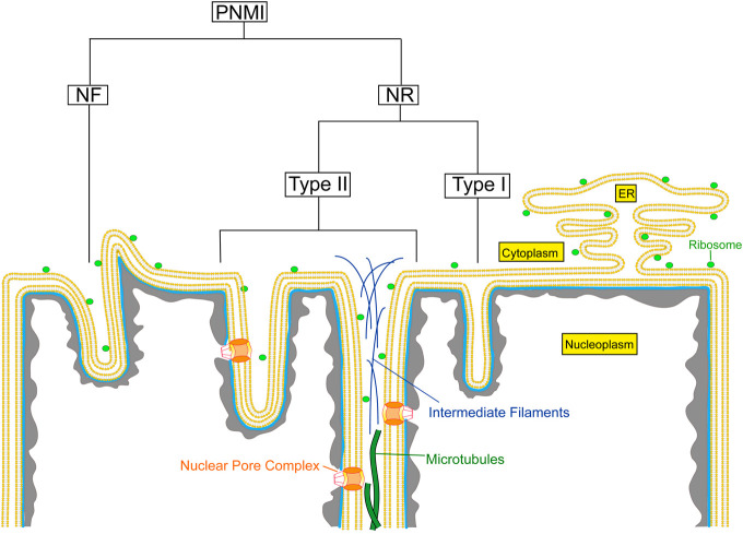
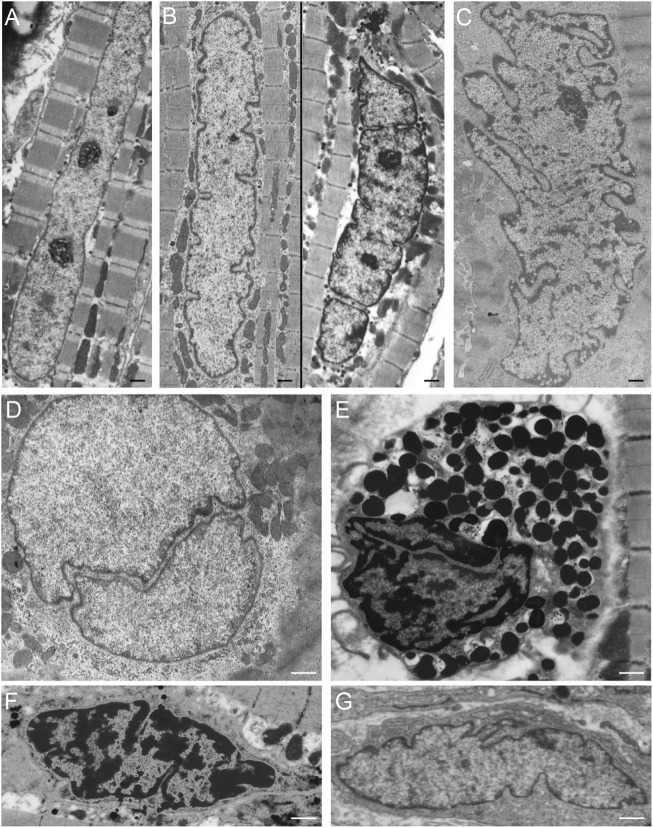
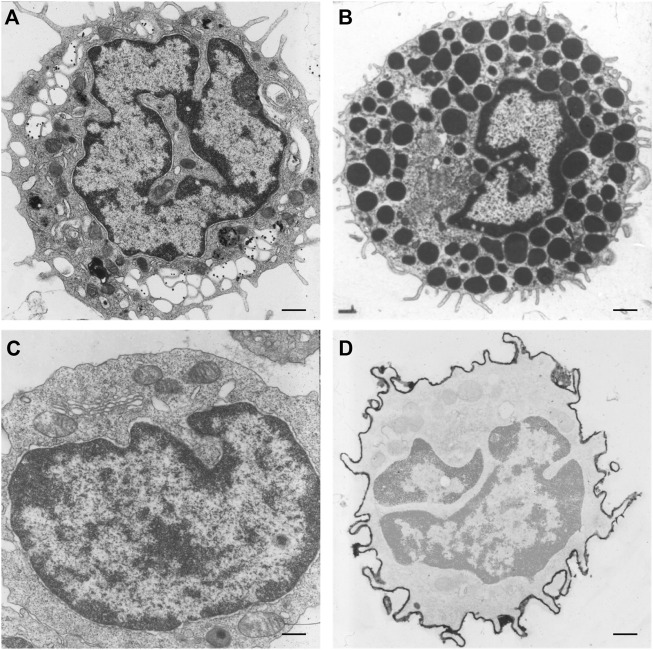
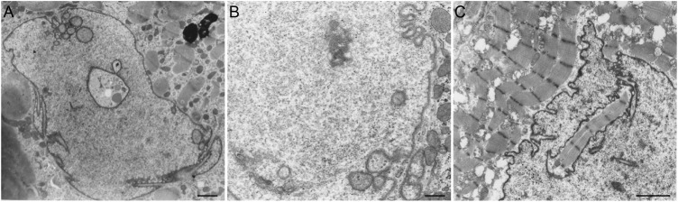
Invaginations of the nuclear membrane occur in different shapes, sizes, and compositions. Part of these pleiomorphic invaginations make up the nucleoplasmic reticulum (NR), while others are merely nuclear folds. We define the NR as tubular invaginations consisting of either both the inner and outer nuclear membrane, or only the inner nuclear membrane. Specifically, invaginations of both the inner and outer nuclear membrane are also called type II NR, while those of only the inner nuclear membrane are defined as type I NR. The formation and structure of the NR is determined by proteins associated to the nuclear membrane, which induce a high membrane curvature leading to tubular invaginations. Here we review and discuss the current knowledge of nuclear invaginations and the NR in particular. An increase in tubular invaginations of the nuclear envelope is associated with several pathologies, such as laminopathies, cancer, (reversible) heart failure, and Alzheimer's disease. Furthermore, viruses can induce both type I and II NR. In laminopathies, the amount of A-type lamins throughout the nucleus is generally decreased or the organization of lamins or lamin-associated proteins is disturbed. Also, lamin overexpression or modulation of lamin farnesylation status impacts NR formation, confirming the importance of lamin processing in NR formation. Virus infections reorganize the nuclear lamina (de)phosphorylation of lamins, leading to an uneven thickness of the nuclear lamina and in turn lobulation of the nuclear membrane and the formation of invaginations of the inner nuclear membrane. Since most studies on the NR have been performed with cell cultures, we present additional proof for the existence of these structures , focusing on a variety of differentiated cardiovascular and hematopoietic cells. Furthermore, we substantiate the knowledge of the lamin composition of the NR by super-resolution images of the lamin A/C and B1 organization. Finally, we further highlight the essential role of lamins in NR formation by demonstrating that (over)expression of lamins can induce aberrant NR structures.
核膜内陷呈现出不同的形状、大小和组成。这些多形性内陷的一部分构成了核质网(NR),而其他的则仅仅是核褶皱。我们将NR定义为由内核膜和外核膜两者,或仅由内核膜组成的管状内陷。具体而言,内核膜和外核膜的内陷也被称为II型NR,而仅内核膜的内陷则被定义为I型NR。NR的形成和结构由与核膜相关的蛋白质决定,这些蛋白质会诱导高膜曲率从而导致管状内陷。在此,我们回顾并讨论关于核内陷尤其是NR的当前知识。核膜管状内陷的增加与多种病理状况相关,如核纤层蛋白病、癌症、(可逆性)心力衰竭和阿尔茨海默病。此外,病毒可诱导I型和II型NR。在核纤层蛋白病中,整个细胞核中A型核纤层蛋白的数量通常会减少,或者核纤层蛋白或核纤层相关蛋白的组织会受到干扰。同样,核纤层蛋白的过表达或法尼基化状态的调节会影响NR的形成,证实了核纤层蛋白加工在NR形成中的重要性。病毒感染会使核纤层蛋白发生(去)磷酸化,从而重组核纤层,导致核纤层厚度不均,进而使核膜形成叶状以及内核膜内陷的形成。由于大多数关于NR的研究是在细胞培养中进行的,我们提供了这些结构存在的额外证据,重点关注各种分化的心血管和造血细胞。此外,我们通过核纤层蛋白A/C和B1组织的超分辨率图像,证实了NR的核纤层蛋白组成的知识。最后,我们通过证明核纤层蛋白的(过)表达可诱导异常的NR结构,进一步强调了核纤层蛋白在NR形成中的关键作用。