Fraunhofer Institute for Toxicology and Experimental Medicine, Hanover, Germany.
Front Public Health. 2022 Jun 21;10:909247. doi: 10.3389/fpubh.2022.909247. eCollection 2022.
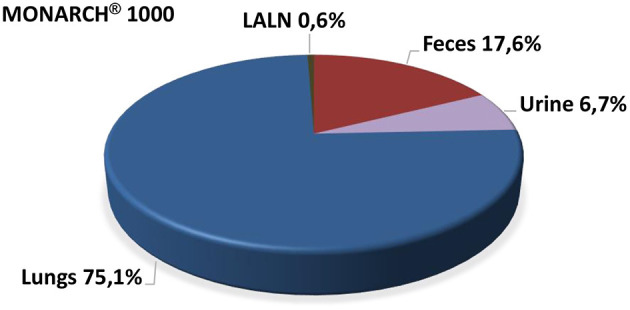
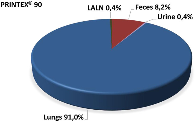
Various synthetic powders with primary particle sizes at the nanoscale and a high commercial impact have been studied using Wistar rats. The test materials were metal oxides, i.e., TiO, ZnO and amorphous silica, and carbon black (technical soot). Dosing schemes were in the regular ranges typically used in subacute rat studies to simulate occupational exposure scenarios (mg range). Nanoscaled particle agglomerates have the potential to disintegrate and translocate as individual nanoparticles to remote locations following deposition in the lungs. The toxicokinetic fate of metal oxides post-inhalation in lungs/organs was investigated (i) by chemical analysis of the retained particulate/dissolved matter and (ii) by visualization of particles in various remote organs using transmission electron microscopy (TEM). The three titanium dioxides (NM-103, NM-104, NM-105; JRC coding) showed a very slow dissolution in lung fluids. In contrast, the coated ZnO (NM-111) dissolved quickly and was eliminated from the body within approximately 1 day. The precipitated amorphous silica (NM-200) showed a partial dissolution. Chemical analysis in lungs (particulate and soluble TiO) and in remote organs (liver and brain) showed a small solubility effect under physiological conditions. The translocation to remote organs was negligible. This confirms that for poorly soluble TiO particles there was no considerable translocation to the liver and brain. The chemical analysis of zinc demonstrated a very rapid dissolution of ZnO particles after deposition in the lungs. Statistically significant increases in Zn levels in the lungs were detectable only on day 1 post-exposure (NM-111). Overall, no relevant amounts of increased NM-111 in the ionic or particulate matter were detected in any body compartment. Amorphous silica (NM-200) particles were found in the cytoplasm of intraalveolar macrophages in the lung and the cytoplasm of macrophages in the lung associated lymph node. Interestingly, these particles were found in a few animals of all treatment groups (1, 2.5, and 5 mg/m NM-200) even after 91 days post-exposure. In all other organs of the NM-200 treated animals such as the nasal epithelium, trachea, larynx, liver, spleen, kidney, and mesenteric lymph node no particles were found at any time point investigated. Carbon black was tagged internally ("intrinsically") with a γ tracer (beryllium; half-time: 53.3 days). Due to limited amounts, the test item (0.3 mg per rat lung) was intratracheally instilled into the lungs. This dose avoided a particle overload effect, meaning that the toxicokinetic fate of carbon black could be followed under the approximated physiological conditions of lung clearance. Analysis of the γ labeled carbon black confirmed conclusively that there was no evidence for the translocation of carbon black beyond the lung into the blood or other body compartments. Very small amounts were only detected in lung-associated lymph nodes (LALN). On day 20 post-treatment, upon necropsy, both carbon black samples were practically exclusively found in lungs (75.1% and 91.0%, respectively) and in very small amounts in the lung-associated lymph nodes (LALN), i.e., ~0.5%. In the other organs/tissues, the test item was not significantly detectable. Separation of leukocytes and cell-free supernatant of a bronchoalveolar lavagate by centrifugation revealed that carbon black was completely located in the cell sediment, indicating total engulfment by alveolar macrophages. In conclusion, in occupational settings the nanomaterials titanium dioxide, zinc oxide, amorphous silica, and carbon black acted as microscaled agglomerates, not as individual nanoparticles. They displayed no potential to translocate beyond the lung into the blood compartment. Besides lungs, very small particulate amounts were detected only in LALN. This finding is consistent with the behavior of microscaled poorly soluble particles. Overall, there was no evidence of translocation of the nanomaterials following pulmonary exposures.
各种具有纳米级初级粒径和高商业影响力的合成粉末已在 Wistar 大鼠中进行了研究。测试材料为金属氧化物,即 TiO、ZnO 和无定形二氧化硅以及炭黑(工业炭黑)。剂量方案处于模拟职业暴露情况的亚急性大鼠研究中通常使用的常规范围内(mg 范围)。纳米级颗粒团聚体有可能在肺部沉积后作为单个纳米颗粒解体并转移到远处。研究了吸入肺部后金属氧化物的毒代动力学命运(i)通过对保留的颗粒/溶解物质进行化学分析,(ii)通过透射电子显微镜(TEM)在各种远程器官中观察颗粒进行。三种二氧化钛(NM-103、NM-104、NM-105;JRC 编码)在肺液中溶解非常缓慢。相比之下,涂层氧化锌(NM-111)迅速溶解,并在大约 1 天内从体内消除。沉淀的无定形二氧化硅(NM-200)显示出部分溶解。肺部(颗粒和可溶性 TiO)和远程器官(肝脏和大脑)中的化学分析表明,在生理条件下,溶解度效应较小。向远程器官的转移可以忽略不计。这证实了对于难溶性 TiO 颗粒,没有向肝脏和大脑的大量转移。锌的化学分析表明,氧化锌颗粒在肺部沉积后迅速溶解。仅在暴露后第 1 天(NM-111)可检测到肺部中 Zn 水平的统计学显着增加。总体而言,在任何身体部位都没有检测到增加的 NM-111 的离子或颗粒物质。无定形二氧化硅(NM-200)颗粒在肺中的肺泡巨噬细胞的细胞质中和与肺相关的淋巴结中的巨噬细胞的细胞质中被发现。有趣的是,即使在暴露后 91 天,所有处理组(1、2.5 和 5mg/m NM-200)的一些动物中也发现了这些颗粒。在 NM-200 处理动物的所有其他器官中,如鼻腔上皮、气管、喉、肝、脾、肾和肠系膜淋巴结,在任何时间点均未发现颗粒。炭黑在内部(“固有地”)标记有γ示踪剂(铍;半衰期:53.3 天)。由于数量有限,将测试物品(每只大鼠肺 0.3mg)通过气管内滴注到肺部。该剂量避免了颗粒过载效应,这意味着可以在近似的肺部清除生理条件下跟踪炭黑的毒代动力学命运。γ 标记的炭黑分析明确证实,没有证据表明炭黑从肺部转移到血液或其他身体部位。仅在肺相关淋巴结(LALN)中检测到非常少量的物质。在治疗后第 20 天,解剖时,两个炭黑样本实际上仅在肺部(分别为 75.1%和 91.0%)和肺相关淋巴结(LALN)中以非常小的量被发现,即~0.5%。在其他器官/组织中,未显著检测到测试物品。通过离心分离支气管肺泡灌洗液中的白细胞和无细胞上清液表明,炭黑完全位于细胞沉积物中,表明肺泡巨噬细胞完全吞噬了它。总之,在职业环境中,纳米材料二氧化钛、氧化锌、无定形二氧化硅和炭黑表现为微尺度团聚体,而不是单个纳米颗粒。它们没有向肺部以外的血液部位转移的潜力。除了肺部,仅在 LALN 中检测到非常小的颗粒量。这一发现与微尺度难溶性颗粒的行为一致。总体而言,没有证据表明肺部暴露后会发生纳米材料的转移。