• 文献检索
  • 文档翻译
  • 深度研究
  • 学术资讯
  • Suppr Zotero 插件Zotero 插件
  • 邀请有礼
  • 套餐&价格
  • 历史记录
应用&插件
Suppr Zotero 插件Zotero 插件浏览器插件Mac 客户端Windows 客户端微信小程序
定价
高级版会员购买积分包购买API积分包
服务
文献检索文档翻译深度研究API 文档MCP 服务
关于我们
关于 Suppr公司介绍联系我们用户协议隐私条款
关注我们

Suppr 超能文献

核心技术专利:CN118964589B侵权必究
粤ICP备2023148730 号-1Suppr @ 2026

文献检索

告别复杂PubMed语法,用中文像聊天一样搜索,搜遍4000万医学文献。AI智能推荐,让科研检索更轻松。

立即免费搜索

文件翻译

保留排版,准确专业,支持PDF/Word/PPT等文件格式,支持 12+语言互译。

免费翻译文档

深度研究

AI帮你快速写综述,25分钟生成高质量综述,智能提取关键信息,辅助科研写作。

立即免费体验

负载碘化油的荧光介孔二氧化硅包覆氧化铁纳米粒子用于磁共振成像/计算机断层扫描/荧光三模态成像。

Iodinated oil-loaded, fluorescent mesoporous silica-coated iron oxide nanoparticles for magnetic resonance imaging/computed tomography/fluorescence trimodal imaging.

作者信息

Xue Sihan, Wang Yao, Wang Mengxing, Zhang Lu, Du Xiaoxia, Gu Hongchen, Zhang Chunfu

机构信息

School of Biomedical Engineering and Med-X Research Institute, Shanghai Jiao Tong University, Shanghai, People's Republic of China.

Shanghai Key Laboratory of Magnetic Resonance, Department of Physics, East China Normal University, Shanghai, People's Republic of China.

出版信息

Int J Nanomedicine. 2014 May 21;9:2527-38. doi: 10.2147/IJN.S59754. eCollection 2014.

DOI:10.2147/IJN.S59754
PMID:24904212
原文链接:https://pmc.ncbi.nlm.nih.gov/articles/PMC4039419/
Abstract

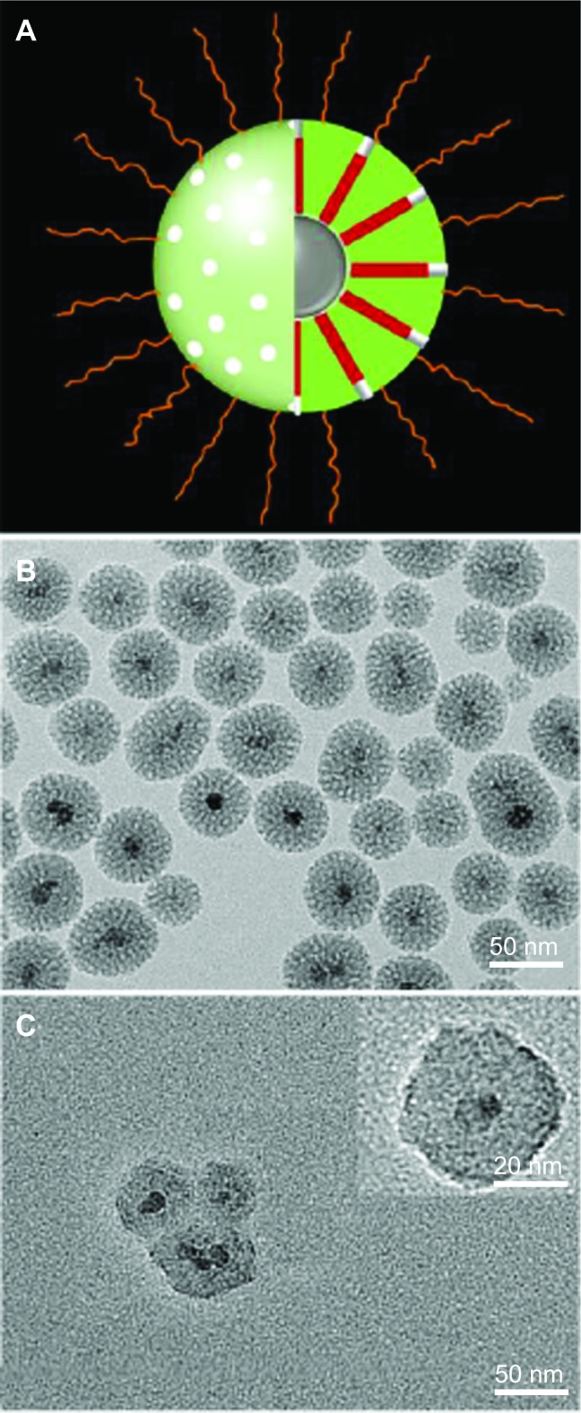
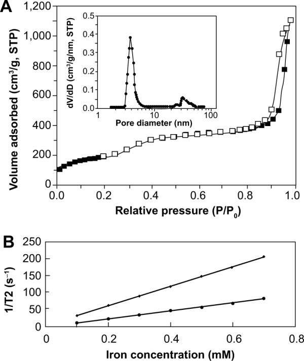
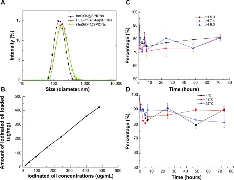
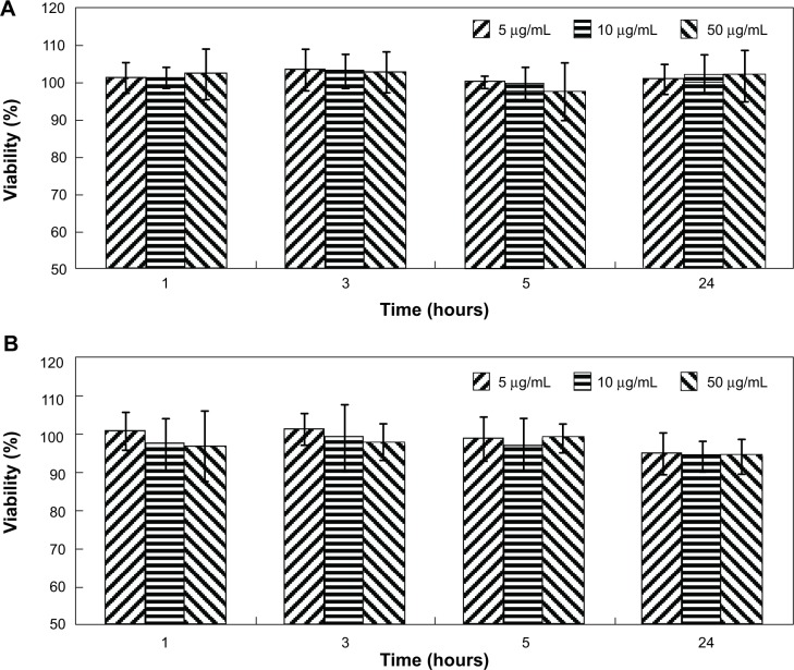
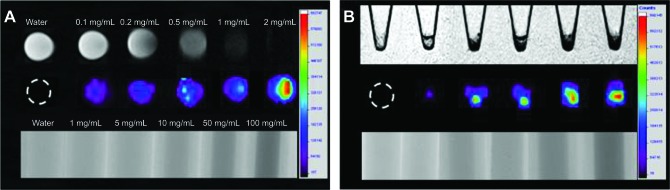
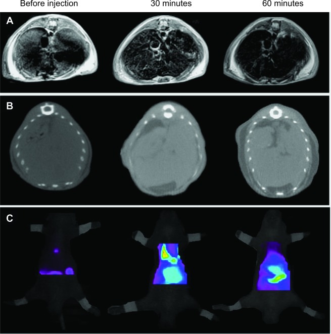
In this study, a novel magnetic resonance imaging (MRI)/computed tomography (CT)/fluorescence trifunctional probe was prepared by loading iodinated oil into fluorescent mesoporous silica-coated superparamagnetic iron oxide nanoparticles (i-fmSiO4@SPIONs). Fluorescent mesoporous silica-coated superparamagnetic iron oxide nanoparticles (fmSiO4@SPIONs) were prepared by growing fluorescent dye-doped silica onto superparamagnetic iron oxide nanoparticles (SPIONs) directed by a cetyltrimethylammonium bromide template. As prepared, fmSiO4@SPIONs had a uniform size, a large surface area, and a large pore volume, which demonstrated high efficiency for iodinated oil loading. Iodinated oil loading did not change the sizes of fmSiO4@SPIONs, but they reduced the MRI T2 relaxivity (r2) markedly. I-fmSiO4@SPIONs were stable in their physical condition and did not demonstrate cytotoxic effects under the conditions investigated. In vitro studies indicated that the contrast enhancement of MRI and CT, and the fluorescence signal intensity of i-fmSiO4@SPION aqueous suspensions and macrophages, were intensified with increased i-fmSiO4@SPION concentrations in suspension and cell culture media. Moreover, for the in vivo study, the accumulation of i-fmSiO4@SPIONs in the liver could also be detected by MRI, CT, and fluorescence imaging. Our study demonstrated that i-fmSiO4@SPIONs had great potential for MRI/CT/fluorescence trimodal imaging.

摘要

在本研究中,通过将碘化油负载到荧光介孔二氧化硅包覆的超顺磁性氧化铁纳米颗粒(i-fmSiO4@SPIONs)中,制备了一种新型的磁共振成像(MRI)/计算机断层扫描(CT)/荧光三功能探针。荧光介孔二氧化硅包覆的超顺磁性氧化铁纳米颗粒(fmSiO4@SPIONs)是通过在十六烷基三甲基溴化铵模板的引导下,将掺杂荧光染料的二氧化硅生长在超顺磁性氧化铁纳米颗粒(SPIONs)上制备而成。所制备的fmSiO4@SPIONs具有均匀的尺寸、较大的表面积和较大的孔体积,这表明其对碘化油的负载效率较高。负载碘化油并未改变fmSiO4@SPIONs的尺寸,但显著降低了MRI的T2弛豫率(r2)。i-fmSiO4@SPIONs在其物理状态下是稳定的,并且在所研究的条件下未表现出细胞毒性作用。体外研究表明,随着悬浮液和细胞培养基中i-fmSiO4@SPION浓度的增加,MRI和CT的对比增强以及i-fmSiO4@SPION水悬浮液和巨噬细胞的荧光信号强度均增强。此外,在体内研究中,也可通过MRI、CT和荧光成像检测到i-fmSiO4@SPIONs在肝脏中的蓄积。我们的研究表明,i-fmSiO4@SPIONs在MRI/CT/荧光三模态成像方面具有巨大潜力。

https://cdn.ncbi.nlm.nih.gov/pmc/blobs/6bc8/4039419/066e00ed1492/ijn-9-2527Fig7.jpg
https://cdn.ncbi.nlm.nih.gov/pmc/blobs/6bc8/4039419/f523b92c88cc/ijn-9-2527Fig1.jpg
https://cdn.ncbi.nlm.nih.gov/pmc/blobs/6bc8/4039419/e45cd246b38e/ijn-9-2527Fig2.jpg
https://cdn.ncbi.nlm.nih.gov/pmc/blobs/6bc8/4039419/5e681c3ce90a/ijn-9-2527Fig3.jpg
https://cdn.ncbi.nlm.nih.gov/pmc/blobs/6bc8/4039419/a07097c7ee3e/ijn-9-2527Fig4.jpg
https://cdn.ncbi.nlm.nih.gov/pmc/blobs/6bc8/4039419/cb9ea50f50da/ijn-9-2527Fig5.jpg
https://cdn.ncbi.nlm.nih.gov/pmc/blobs/6bc8/4039419/cccde2329cac/ijn-9-2527Fig6.jpg
https://cdn.ncbi.nlm.nih.gov/pmc/blobs/6bc8/4039419/066e00ed1492/ijn-9-2527Fig7.jpg

相似文献

1
Iodinated oil-loaded, fluorescent mesoporous silica-coated iron oxide nanoparticles for magnetic resonance imaging/computed tomography/fluorescence trimodal imaging.负载碘化油的荧光介孔二氧化硅包覆氧化铁纳米粒子用于磁共振成像/计算机断层扫描/荧光三模态成像。
Int J Nanomedicine. 2014 May 21;9:2527-38. doi: 10.2147/IJN.S59754. eCollection 2014.
2
High MRI performance fluorescent mesoporous silica-coated magnetic nanoparticles for tracking neural progenitor cells in an ischemic mouse model.用于在缺血性小鼠模型中追踪神经祖细胞的高 MRI 性能荧光介孔硅涂层磁性纳米颗粒。
Nanoscale. 2013 May 21;5(10):4506-16. doi: 10.1039/c3nr00119a.
3
Surface engineered antifouling optomagnetic SPIONs for bimodal targeted imaging of pancreatic cancer cells.表面工程化的抗污光磁 SPIONs 用于胰腺癌双模态靶向成像。
Int J Nanomedicine. 2014 Mar 27;9:1601-15. doi: 10.2147/IJN.S58334. eCollection 2014.
4
Targeted Molecular Iron Oxide Contrast Agents for Imaging Atherosclerotic Plaque.靶向分子氧化铁对比剂用于动脉粥样硬化斑块成像。
Nanotheranostics. 2020 May 30;4(4):184-194. doi: 10.7150/ntno.44712. eCollection 2020.
5
Magnetite- and Iodine-Containing Nanoemulsion as a Dual Modal Contrast Agent for X-ray/Magnetic Resonance Imaging.载磁铁矿和碘的纳米乳剂作为 X 射线/磁共振成像的双模态对比剂。
ACS Appl Mater Interfaces. 2019 Jan 9;11(1):403-416. doi: 10.1021/acsami.8b19517. Epub 2018 Dec 26.
6
One-pot facile synthesis of PEGylated superparamagnetic iron oxide nanoparticles for MRI contrast enhancement.一锅法简便合成用于 MRI 对比增强的 PEG 化超顺磁性氧化铁纳米颗粒。
Mater Sci Eng C Mater Biol Appl. 2014 Aug 1;41:161-7. doi: 10.1016/j.msec.2014.04.041. Epub 2014 Apr 28.
7
Tunable and noncytotoxic PET/SPECT-MRI multimodality imaging probes using colloidally stable ligand-free superparamagnetic iron oxide nanoparticles.使用胶体稳定的无配体超顺磁性氧化铁纳米颗粒的可调节且无细胞毒性的正电子发射断层扫描/单光子发射计算机断层扫描-磁共振成像多模态成像探针。
Int J Nanomedicine. 2017 Jan 27;12:899-909. doi: 10.2147/IJN.S127171. eCollection 2017.
8
Facile Synthesis of Gold Nanospheres Modified by Positively Charged Mesoporous Silica, Loaded with Near-Infrared Fluorescent Dye, for in Vivo X-ray Computed Tomography and Fluorescence Dual Mode Imaging.简便合成带正电荷的介孔二氧化硅修饰的金纳米球,负载近红外荧光染料,用于体内X射线计算机断层扫描和荧光双模式成像。
ACS Appl Mater Interfaces. 2015 Aug 12;7(31):17287-97. doi: 10.1021/acsami.5b04359. Epub 2015 Jul 30.
9
Preliminary Study of MR and Fluorescence Dual-mode Imaging: Combined Macrophage-Targeted and Superparamagnetic Polymeric Micelles.磁共振与荧光双模成像的初步研究:巨噬细胞靶向与超顺磁性聚合物胶束联合
Int J Med Sci. 2018 Jan 1;15(2):129-141. doi: 10.7150/ijms.21610. eCollection 2018.
10
Clustering superparamagnetic iron oxide nanoparticles produces organ-targeted high-contrast magnetic resonance images.超顺磁性氧化铁纳米粒子的聚类生成靶向器官的高对比度磁共振图像。
Nanomedicine (Lond). 2019 May;14(9):1135-1152. doi: 10.2217/nnm-2018-0370. Epub 2019 May 3.

引用本文的文献

1
Magnetic Resonance Imaging and X-Ray Imaging Properties of Ultrasmall Lanthanide Oxide (Ln = Eu, Gd, and Tb) Nanoparticles Synthesized via Thermal Decomposition.通过热分解法合成的超小镧系氧化物(Ln = Eu、Gd和Tb)纳米粒子的磁共振成像和X射线成像特性
Molecules. 2025 Jun 9;30(12):2519. doi: 10.3390/molecules30122519.
2
Nanomaterial-based CT contrast agents and their applications in image-guided therapy.基于纳米材料的 CT 对比剂及其在影像引导治疗中的应用。
Theranostics. 2023 Jan 1;13(2):483-509. doi: 10.7150/thno.79625. eCollection 2023.
3
Nanoarchitectured prototypes of mesoporous silica nanoparticles for innovative biomedical applications.

本文引用的文献

1
In vivo tomographic imaging with fluorescence and MRI using tumor-targeted dual-labeled nanoparticles.肿瘤靶向双标记纳米粒子的荧光和 MRI 的体内断层成像。
Int J Nanomedicine. 2014;9:33-41. doi: 10.2147/IJN.S52492. Epub 2013 Dec 16.
2
Recent progress in rare earth micro/nanocrystals: soft chemical synthesis, luminescent properties, and biomedical applications.稀土微纳晶体的最新进展:软化学合成、发光特性及生物医学应用
Chem Rev. 2014 Feb 26;114(4):2343-89. doi: 10.1021/cr4001594. Epub 2013 Dec 18.
3
Upconverting and NIR emitting rare earth based nanostructures for NIR-bioimaging.
介孔硅纳米粒子的纳米结构原型用于创新的生物医学应用。
J Nanobiotechnology. 2022 Mar 12;20(1):126. doi: 10.1186/s12951-022-01315-x.
4
Metal Oxide Nanoparticles: Evidence of Adverse Effects on the Male Reproductive System.金属氧化物纳米颗粒:对男性生殖系统不良影响的证据。
Int J Mol Sci. 2021 Jul 28;22(15):8061. doi: 10.3390/ijms22158061.
5
Imaging Constructs: The Rise of Iron Oxide Nanoparticles.成像构建体:氧化铁纳米粒子的崛起。
Molecules. 2021 Jun 5;26(11):3437. doi: 10.3390/molecules26113437.
6
Magnetic Nanoparticles-A Multifunctional Potential Agent for Diagnosis and Therapy.磁性纳米粒子——一种用于诊断和治疗的多功能潜在试剂。
Cancers (Basel). 2021 May 5;13(9):2213. doi: 10.3390/cancers13092213.
7
Tumor Chemo-Radiotherapy with Rod-Shaped and Spherical Gold Nano Probes: Shape and Active Targeting Both Matter.棒状和球形金纳米探针的肿瘤化放疗:形状和主动靶向都很重要。
Theranostics. 2019 Mar 16;9(7):1893-1908. doi: 10.7150/thno.30523. eCollection 2019.
8
Biomedical Imaging: Principles, Technologies, Clinical Aspects, Contrast Agents, Limitations and Future Trends in Nanomedicines.生物医学成像:原理、技术、临床方面、对比剂、纳米医学的局限性和未来趋势。
Pharm Res. 2019 Apr 3;36(6):78. doi: 10.1007/s11095-019-2608-5.
9
Theragnostic potentials of core/shell mesoporous silica nanostructures.核/壳介孔二氧化硅纳米结构的诊疗潜力。
Nanotheranostics. 2019 Jan 1;3(1):1-40. doi: 10.7150/ntno.27877. eCollection 2019.
10
toxicity evaluation of silica-coated iron oxide nanoparticles in human SHSY5Y neuronal cells.二氧化硅包覆的氧化铁纳米颗粒在人SHSY5Y神经细胞中的毒性评估
Toxicol Res (Camb). 2015 Oct 23;5(1):235-247. doi: 10.1039/c5tx00206k. eCollection 2016 Jan 1.
上转换和近红外发射稀土基纳米结构用于近红外生物成像。
Nanoscale. 2013 Dec 7;5(23):11339-61. doi: 10.1039/c3nr02286b. Epub 2013 Aug 12.
4
Multifunctional upconversion mesoporous silica nanostructures for dual modal imaging and in vivo drug delivery.多功能上转换介孔硅纳米结构用于双模式成像和体内药物输送。
Small. 2013 Dec 20;9(24):4150-9. doi: 10.1002/smll.201301093. Epub 2013 Jul 11.
5
Extensive evaluations of the cytotoxic effects of gold nanoparticles.对金纳米颗粒细胞毒性作用的广泛评估。
Biochim Biophys Acta. 2013 Oct;1830(10):4960-73. doi: 10.1016/j.bbagen.2013.06.025. Epub 2013 Jun 27.
6
Magnetic nanoparticles as contrast agents in the diagnosis and treatment of cancer.磁性纳米颗粒作为癌症诊断和治疗的对比剂。
Chem Soc Rev. 2013 Oct 7;42(19):7816-33. doi: 10.1039/c3cs60149h. Epub 2013 Jun 21.
7
Uptake kinetics and nanotoxicity of silica nanoparticles are cell type dependent.硅纳米颗粒的摄取动力学和纳米毒性取决于细胞类型。
Small. 2013 Dec 9;9(23):3970-80, 3906. doi: 10.1002/smll.201301004. Epub 2013 May 17.
8
High MRI performance fluorescent mesoporous silica-coated magnetic nanoparticles for tracking neural progenitor cells in an ischemic mouse model.用于在缺血性小鼠模型中追踪神经祖细胞的高 MRI 性能荧光介孔硅涂层磁性纳米颗粒。
Nanoscale. 2013 May 21;5(10):4506-16. doi: 10.1039/c3nr00119a.
9
Targeted CT/MR dual mode imaging of tumors using multifunctional dendrimer-entrapped gold nanoparticles.利用多功能树枝状大分子包裹的金纳米粒子进行肿瘤的靶向 CT/MR 双模式成像。
Biomaterials. 2013 Jul;34(21):5200-9. doi: 10.1016/j.biomaterials.2013.03.009. Epub 2013 Apr 10.
10
A concise review of magnetic resonance molecular imaging of tumor angiogenesis by targeting integrin αvβ3 with magnetic probes.针对整合素 αvβ3 的磁探针进行肿瘤血管生成的磁共振分子成像的简明综述。
Int J Nanomedicine. 2013;8:1083-93. doi: 10.2147/IJN.S39880. Epub 2013 Mar 14.