• 文献检索
  • 文档翻译
  • 深度研究
  • 学术资讯
  • Suppr Zotero 插件Zotero 插件
  • 邀请有礼
  • 套餐&价格
  • 历史记录
应用&插件
Suppr Zotero 插件Zotero 插件浏览器插件Mac 客户端Windows 客户端微信小程序
定价
高级版会员购买积分包购买API积分包
服务
文献检索文档翻译深度研究API 文档MCP 服务
关于我们
关于 Suppr公司介绍联系我们用户协议隐私条款
关注我们

Suppr 超能文献

核心技术专利:CN118964589B侵权必究
粤ICP备2023148730 号-1Suppr @ 2026

文献检索

告别复杂PubMed语法,用中文像聊天一样搜索,搜遍4000万医学文献。AI智能推荐,让科研检索更轻松。

立即免费搜索

文件翻译

保留排版,准确专业,支持PDF/Word/PPT等文件格式,支持 12+语言互译。

免费翻译文档

深度研究

AI帮你快速写综述,25分钟生成高质量综述,智能提取关键信息,辅助科研写作。

立即免费体验

准确的基于图像的侧脑室脑脊液容量计算。

Accurate image-based CSF volume calculation of the lateral ventricles.

机构信息

Science Based Platforms LLC, R&D, 604 Beach CT, Fort Pierce, 34950, USA.

GYM Group SA, R&D, Carrera 78A 6-58, Cali, Valle del Cauca, 76001, Colombia.

出版信息

Sci Rep. 2022 Jul 15;12(1):12115. doi: 10.1038/s41598-022-15995-w.

DOI:10.1038/s41598-022-15995-w
PMID:35840587
原文链接:https://pmc.ncbi.nlm.nih.gov/articles/PMC9287564/
Abstract

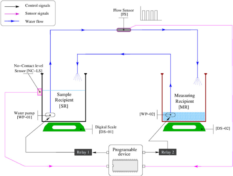
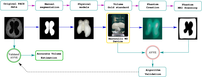
The size/volume of the brain's ventricles is essential in diagnosing and treating many neurological disorders, with various forms of hydrocephalus being some of the most common. Initial ventricular size and changes, if any, in response to disease progression or therapeutic intervention are monitored by serial imaging methods. Significant variance in ventricular size is readily noted, but small incremental changes can be challenging to appreciate. We have previously reported using artificial intelligence to determine ventricular volume. The values obtained were compared with those calculated using the inaccurate manual segmentation as the "gold standard". This document introduces a strategy to measure ventricular volumes where manual segmentation is not employed to validate the estimations. Instead, we created 3D printed models that mimic the lateral ventricles and measured those 3D models' volume with a tuned water displacement device. The 3D models are placed in a gel and taken to the magnetic resonance scanner. Images extracted from the phantoms are fed to an artificial intelligence-based algorithm. The volumes yielded by the automation must equal those yielded by water displacement to assert validation. Then, we provide certified volumes for subjects in the age range (1-114) months old and two hydrocephalus patients.

摘要

脑室内径的大小/体积对于诊断和治疗许多神经疾病至关重要,其中各种类型的脑积水是最常见的疾病之一。通过连续的影像学方法来监测初始脑室大小以及疾病进展或治疗干预后的任何变化。很容易注意到脑室大小的显著差异,但小的增量变化可能难以察觉。我们之前曾报道过使用人工智能来确定脑室体积。将获得的值与使用不准确的手动分割作为“金标准”计算的值进行比较。本文介绍了一种测量脑室体积的策略,其中不采用手动分割来验证估计值。相反,我们创建了模拟侧脑室的 3D 打印模型,并使用经过调整的水置换设备来测量这些 3D 模型的体积。将 3D 模型放置在凝胶中并带到磁共振扫描仪中。从模型中提取图像并输入基于人工智能的算法。自动化生成的体积必须等于水置换的体积,以确保验证。然后,我们为年龄在(1-114)个月之间的受试者和两名脑积水患者提供认证的体积。

https://cdn.ncbi.nlm.nih.gov/pmc/blobs/1013/9287564/1e8f061c147a/41598_2022_15995_Fig5_HTML.jpg
https://cdn.ncbi.nlm.nih.gov/pmc/blobs/1013/9287564/6744d45ec01c/41598_2022_15995_Fig1_HTML.jpg
https://cdn.ncbi.nlm.nih.gov/pmc/blobs/1013/9287564/54697cb7f625/41598_2022_15995_Fig2_HTML.jpg
https://cdn.ncbi.nlm.nih.gov/pmc/blobs/1013/9287564/80c8d48a8762/41598_2022_15995_Fig3_HTML.jpg
https://cdn.ncbi.nlm.nih.gov/pmc/blobs/1013/9287564/f8307aecff9a/41598_2022_15995_Fig4_HTML.jpg
https://cdn.ncbi.nlm.nih.gov/pmc/blobs/1013/9287564/1e8f061c147a/41598_2022_15995_Fig5_HTML.jpg

相似文献

1
Accurate image-based CSF volume calculation of the lateral ventricles.准确的基于图像的侧脑室脑脊液容量计算。
Sci Rep. 2022 Jul 15;12(1):12115. doi: 10.1038/s41598-022-15995-w.
2
Eliminating the need for manual segmentation to determine size and volume from MRI. A proof of concept on segmenting the lateral ventricles.消除手动分割以从 MRI 确定大小和体积的需求。对侧脑室分割的概念验证。
PLoS One. 2023 May 11;18(5):e0285414. doi: 10.1371/journal.pone.0285414. eCollection 2023.
3
Artificial intelligence for automatic cerebral ventricle segmentation and volume calculation: a clinical tool for the evaluation of pediatric hydrocephalus.人工智能在自动脑室内层分割和容量计算中的应用:小儿脑积水评估的临床工具。
J Neurosurg Pediatr. 2020 Dec 1;27(2):131-138. doi: 10.3171/2020.6.PEDS20251. Print 2021 Feb 1.
4
Automated Lateral Ventricular and Cranial Vault Volume Measurements in 13,851 Patients Using Deep Learning Algorithms.使用深度学习算法对13851例患者进行侧脑室和颅腔容积的自动测量
World Neurosurg. 2021 Apr;148:e363-e373. doi: 10.1016/j.wneu.2020.12.148. Epub 2021 Jan 6.
5
Volume estimation of brain ventricles using Cavalieri's principle and Atlas-based methods in Alzheimer disease: Consistency between methods.应用卡瓦列里原理和基于图谱的方法对阿尔茨海默病患者脑室内容量进行评估:方法间的一致性。
J Clin Neurosci. 2020 Aug;78:333-338. doi: 10.1016/j.jocn.2020.04.092. Epub 2020 Apr 29.
6
Ventricular Volume Dynamics During the Development of Adult Chronic Communicating Hydrocephalus in a Rodent Model.啮齿动物模型中成年慢性交通性脑积水发展过程中的心室容积动态变化
World Neurosurg. 2018 Dec;120:e1120-e1127. doi: 10.1016/j.wneu.2018.08.241. Epub 2018 Sep 11.
7
Is all "communicating" hydrocephalus really communicating? Prospective study on the value of 3D-constructive interference in steady state sequence at 3T.所有的“交通性”脑积水真的是交通性的吗?3T 三维稳态干扰真实快速成像序列对交通性脑积水的前瞻性研究
AJNR Am J Neuroradiol. 2009 Nov;30(10):1898-906. doi: 10.3174/ajnr.A1726. Epub 2009 Jul 30.
8
Influence of shunt type on ventricular volume changes in children with hydrocephalus.分流类型对脑积水患儿脑室容积变化的影响。
J Neurosurg. 2003 Feb;98(2):277-83. doi: 10.3171/jns.2003.98.2.0277.
9
Magnetic resonance imaging based correlation analysis between calcarine sulcus development and isolated fetal ventriculomegaly.基于磁共振成像的距状沟发育与孤立性胎儿脑室扩大之间的相关性分析。
Congenit Anom (Kyoto). 2017 Mar;57(2):52-56. doi: 10.1111/cga.12197.
10
Acute hydrocephalus secondary to obstruction of the foramen of monro and cerebral aqueduct caused by a choroid plexus cyst in the lateral ventricle. Case report.侧脑室脉络丛囊肿导致室间孔和中脑导水管梗阻继发急性脑积水。病例报告。
J Neurosurg. 2007 Sep;107(3 Suppl):236-9. doi: 10.3171/PED-07/09/236.

引用本文的文献

1
Automatic measurement and reference values setting of intracranial cerebrospinal fluid volume: a large-scale analysis of computed tomography images.颅内脑脊液体积的自动测量与参考值设定:计算机断层扫描图像的大规模分析
Quant Imaging Med Surg. 2025 Jul 1;15(7):6185-6199. doi: 10.21037/qims-2024-2571. Epub 2025 Jun 30.

本文引用的文献

1
Clinical and neuroimaging findings in MOGAD-MRI and OCT.MOGAD-MRI 和 OCT 的临床和神经影像学表现。
Clin Exp Immunol. 2021 Dec;206(3):266-281. doi: 10.1111/cei.13641. Epub 2021 Jul 18.
2
Automatically measuring brain ventricular volume within PACS using artificial intelligence.利用人工智能在 PACS 中自动测量脑室内容积。
PLoS One. 2018 Mar 15;13(3):e0193152. doi: 10.1371/journal.pone.0193152. eCollection 2018.
3
A 3D subject-specific model of the spinal subarachnoid space with anatomically realistic ventral and dorsal spinal cord nerve rootlets.
具有解剖学真实的腹侧和背侧脊髓神经根的脊髓蛛网膜下腔的三维个体化模型。
Fluids Barriers CNS. 2017 Dec 19;14(1):36. doi: 10.1186/s12987-017-0085-y.
4
The cerebrospinal fluid and barriers - anatomic and physiologic considerations.脑脊液与屏障——解剖学和生理学考量
Handb Clin Neurol. 2017;146:21-32. doi: 10.1016/B978-0-12-804279-3.00002-2.
5
Fetal MRI of the central nervous system: State-of-the-art.中枢神经系统的胎儿磁共振成像:最新技术。
Eur J Radiol. 2017 Aug;93:273-283. doi: 10.1016/j.ejrad.2017.06.004. Epub 2017 Jun 17.
6
Neuroimaging in Central Nervous System Infections.中枢神经系统感染中的神经影像学
Curr Neurol Neurosci Rep. 2017 Jun;17(6):49. doi: 10.1007/s11910-017-0756-8.
7
Automated segmentation of MR imaging to determine normative central nervous system cerebrospinal fluid volumes in healthy volunteers.通过磁共振成像自动分割来确定健康志愿者中枢神经系统脑脊液的标准体积。
Clin Imaging. 2017 May-Jun;43:132-135. doi: 10.1016/j.clinimag.2017.02.007. Epub 2017 Mar 2.
8
Quantitative evaluation of image segmentation incorporating medical consideration functions.结合医学考量函数的图像分割定量评估。
Med Phys. 2015 Jun;42(6):3013-23. doi: 10.1118/1.4921067.
9
Right ventricular segmentation in cardiac MRI with moving mesh correspondences.心脏 MRI 中运用活动网格对应技术进行右心室分段。
Comput Med Imaging Graph. 2015 Jul;43:15-25. doi: 10.1016/j.compmedimag.2015.01.004. Epub 2015 Feb 12.
10
A reproducible evaluation of ANTs similarity metric performance in brain image registration.在脑影像配准中重复评估 ANTs 相似性度量性能。
Neuroimage. 2011 Feb 1;54(3):2033-44. doi: 10.1016/j.neuroimage.2010.09.025. Epub 2010 Sep 17.