Department of Endodontics, School of Stomatology, Capital Medical University, Tian Tan Xi Li No.4, Beijing, 100050, People's Republic of China.
Immunology Research Center for Oral and Systemic Health, Beijing Friendship Hospital, Capital Medical University, Beijing, People's Republic of China.
BMC Oral Health. 2022 Jul 16;22(1):290. doi: 10.1186/s12903-022-02327-7.
External root resorption is one of common complications of orthodontic treatment, while internal root resorption is rarely observed, and the difference between pulp and periodontal tissues during orthodontic treatment is still unknown. The purpose of this study was to evaluate the effects of orthodontic forces on histological and cellular changes of the dental pulp and periodontal tissues.
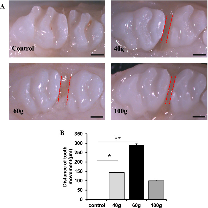
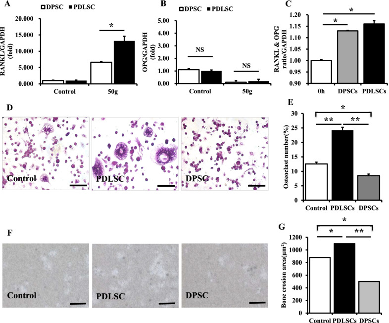
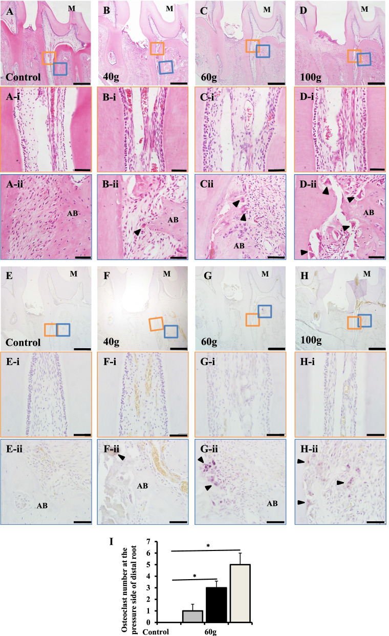
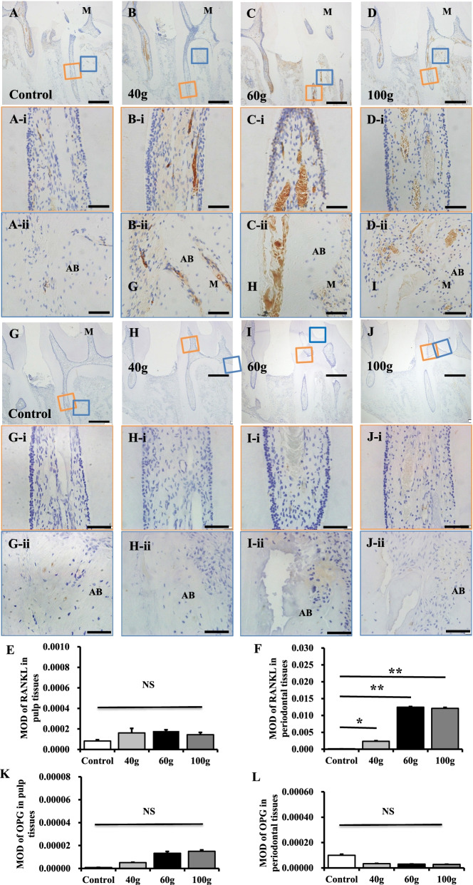
Orthodontic tooth movement model was established in Forty-eight adult male Wistar rats. The distance of orthodontic tooth movement was quantitatively analyzed. The histological changes of pulp and periodontal tissues were performed by hematoxylin-eosin staining, tartrate-resistant acid phosphate staining was used to analyze the changes of osteoclast number, immunohistochemistry analysis and reverse transcription polymerase chain reaction were used to examine the receptor of nuclear factor-κB ligand (RANKL) and osteoprotegerin (OPG) expression. The width of tertiary dentine was quantitatively analyzed. Tartrate-resistant acid phosphate staining and the erosion area of osteo assay surface plate was used to evaluate osteoclast activity.
The orthodontic tooth movement distance increased in a force dependent manner, and reached the peak value when orthodontic force is 60 g. Heavy orthodontic force increased the RANKL expression of periodontal ligament srem cells (PDLSCs) which further activated osteoclasts and resulted in external root resorption, while the RANKL expression of dental pulp stem cells (DPSCs) was relatively low to activate osteoclasts and result in internal root resorption, and the dental pulp tend to form tertiary dentine under orthodontic force stimulation.
Heavy orthodontic forces activated osteoclasts and triggered external root resorption by upregulating RANKL expression in rat periodontal tissues, while there was no significant change of RANKL expression in dental pulp tissue under heavy orthodontic forces, which prevented osteoclast activation and internal root resorption.
牙齿外部吸收是正畸治疗的常见并发症之一,而牙齿内部吸收则很少见,正畸治疗过程中牙髓和牙周组织之间的差异仍不清楚。本研究旨在评估正畸力对牙髓和牙周组织的组织学和细胞变化的影响。
建立 48 只成年雄性 Wistar 大鼠正畸牙移动模型。定量分析正畸牙移动距离。采用苏木精-伊红染色观察牙髓和牙周组织的组织学变化,抗酒石酸酸性磷酸酶染色分析破骨细胞数量的变化,免疫组织化学分析和逆转录聚合酶链反应检测核因子-κB 配体(RANKL)和骨保护素(OPG)的表达。定量分析第三期牙本质的宽度。抗酒石酸酸性磷酸酶染色和破骨细胞表面板侵蚀面积用于评估破骨细胞活性。
正畸牙移动距离呈力依赖性增加,当正畸力为 60g 时达到峰值。大正畸力增加了牙周膜干细胞(PDLSCs)的 RANKL 表达,进而激活破骨细胞,导致牙齿外部吸收,而牙髓干细胞(DPSCs)的 RANKL 表达相对较低,无法激活破骨细胞,导致牙齿内部吸收,且牙髓在正畸力刺激下倾向于形成第三期牙本质。
大正畸力通过上调大鼠牙周组织中 RANKL 的表达激活破骨细胞,引发牙齿外部吸收,而大正畸力下牙髓组织中 RANKL 的表达没有明显变化,防止了破骨细胞的激活和牙齿内部吸收。