Fraunhofer Institute for Digital Medicine MEVIS, Bremen, Germany.
Medical Image Computing Group, University of Bremen, Bremen, Germany.
Sci Rep. 2022 Jul 18;12(1):12262. doi: 10.1038/s41598-022-16388-9.
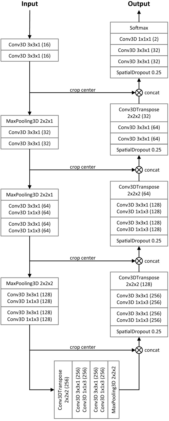
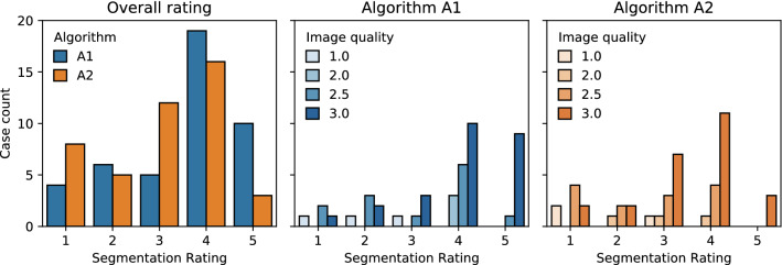
Automatic liver tumor segmentation can facilitate the planning of liver interventions. For diagnosis of hepatocellular carcinoma, dynamic contrast-enhanced MRI (DCE-MRI) can yield a higher sensitivity than contrast-enhanced CT. However, most studies on automatic liver lesion segmentation have focused on CT. In this study, we present a deep learning-based approach for liver tumor segmentation in the late hepatocellular phase of DCE-MRI, using an anisotropic 3D U-Net architecture and a multi-model training strategy. The 3D architecture improves the segmentation performance compared to a previous study using a 2D U-Net (mean Dice 0.70 vs. 0.65). A further significant improvement is achieved by a multi-model training approach (0.74), which is close to the inter-rater agreement (0.78). A qualitative expert rating of the automatically generated contours confirms the benefit of the multi-model training strategy, with 66 % of contours rated as good or very good, compared to only 43 % when performing a single training. The lesion detection performance with a mean F1-score of 0.59 is inferior to human raters (0.76). Overall, this study shows that correctly detected liver lesions in late-phase DCE-MRI data can be automatically segmented with high accuracy, but the detection, in particular of smaller lesions, can still be improved.
自动肝肿瘤分割可以辅助肝介入规划。对于肝细胞癌的诊断,动态对比增强磁共振成像(DCE-MRI)比增强 CT 的灵敏度更高。然而,大多数自动肝病变分割的研究都集中在 CT 上。在这项研究中,我们提出了一种基于深度学习的方法,用于 DCE-MRI 肝细胞晚期肝肿瘤分割,使用各向异性 3D U-Net 架构和多模型训练策略。3D 架构比以前使用 2D U-Net 的研究(平均 Dice 为 0.70 比 0.65)提高了分割性能。通过多模型训练方法(0.74)进一步显著提高,接近组内一致性(0.78)。对自动生成轮廓的定性专家评分证实了多模型训练策略的益处,有 66%的轮廓被评为良好或非常好,而单独训练时只有 43%。平均 F1 得分为 0.59 的病变检测性能不如人类评估者(0.76)。总体而言,这项研究表明,在晚期 DCE-MRI 数据中,可以使用高精度自动分割正确检测到的肝病变,但检测,特别是较小的病变,仍有待提高。