Su Yanlin, Ye Bing, Zeng Lian, Xiong Zekang, Sun Tingfang, Chen Kaifang, Ding Qiuyue, Su Weijie, Jing Xirui, Gao Qing, Huang Guixiong, Wan Yizhou, Yang Xu, Guo Xiaodong
Department of Orthopaedics, Union Hospital, Tongji Medical College, Huazhong University of Science and Technology, Wuhan 430022, China.
Department of Orthopedics, Suizhou Hospital, Hubei University of Medicine, Suizhou 441300, China.
Membranes (Basel). 2022 Jul 20;12(7):719. doi: 10.3390/membranes12070719.
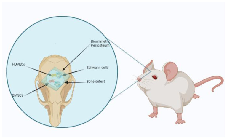
Critical bone defects are a significant problem in clinics. The periosteum plays a vital role in bone regeneration. A tissue-engineered periosteum (TEP) has received increasing attention as a novel strategy for bone defect repairs.
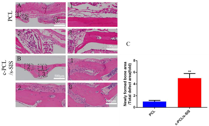
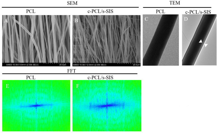
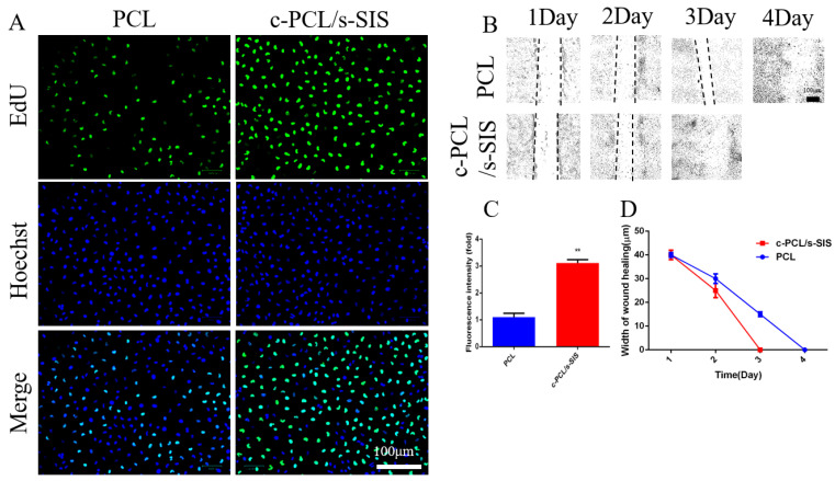
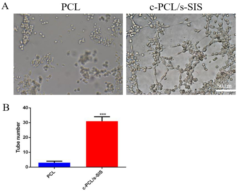
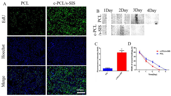
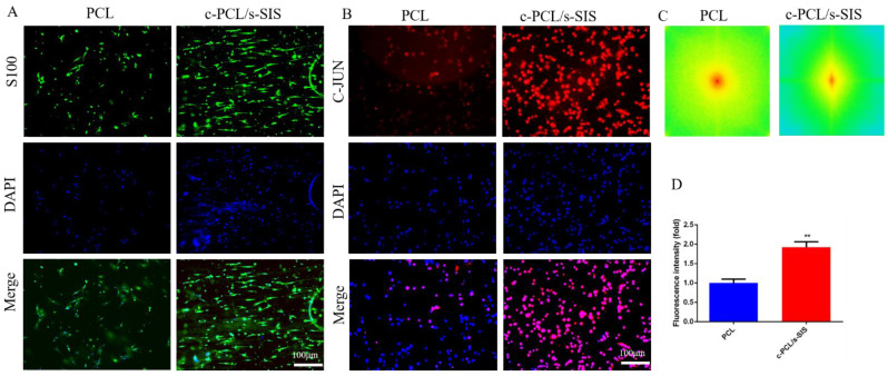
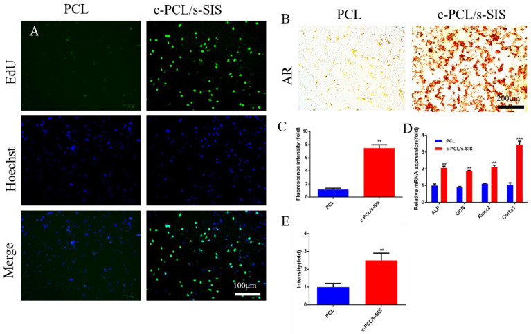
In this experiment, a biomimetic periosteum was fabricated by using coaxial electrospinning technology with decellularized porcine small intestinal submucosa (SIS) as the shell and polycaprolactone (PCL) as the core. In vitro, the effects of the biomimetic periosteum on Schwann cells, vascular endothelial cells, and bone marrow mesenchymal stem cells were detected by a scratch test, an EdU, a tube-forming test, and an osteogenesis test. In vivo, we used HE staining to evaluate the effect of the biomimetic periosteum on bone regeneration.
In vitro experiments showed that the biomimetic periosteum could significantly promote the formation of angiogenesis, osteogenesis, and repaired Schwann cells (SCs). In vivo experiments showed that the biomimetic periosteum could promote the repair of bone defects.
The biomimetic periosteum could simulate the structural function of the periosteum and promote bone repair. This strategy may provide a promising method for the clinical treatment of skull bone defects.
严重骨缺损是临床上的一个重大问题。骨膜在骨再生中起着至关重要的作用。组织工程骨膜(TEP)作为一种骨缺损修复的新策略受到越来越多的关注。
在本实验中,采用同轴静电纺丝技术制备仿生骨膜,以去细胞猪小肠黏膜下层(SIS)为外壳,聚己内酯(PCL)为核心。在体外,通过划痕试验、EdU、管形成试验和成骨试验检测仿生骨膜对雪旺细胞、血管内皮细胞和骨髓间充质干细胞的影响。在体内,我们用苏木精-伊红染色评估仿生骨膜对骨再生的影响。
体外实验表明,仿生骨膜能显著促进血管生成、成骨和雪旺细胞(SCs)的修复。体内实验表明,仿生骨膜能促进骨缺损的修复。
仿生骨膜可模拟骨膜的结构功能,促进骨修复。该策略可能为颅骨缺损的临床治疗提供一种有前景的方法。