Department of Radiology, Washington University School of Medicine, 510 S. Kingshighway Blvd., Campus, Box 8225, St. Louis, MO, 63110, USA.
Program in Quantitative Molecular Therapeutics, Washington University School of Medicine, St. Louis, MO, USA.
Sci Rep. 2022 Jul 29;12(1):13034. doi: 10.1038/s41598-022-17460-0.
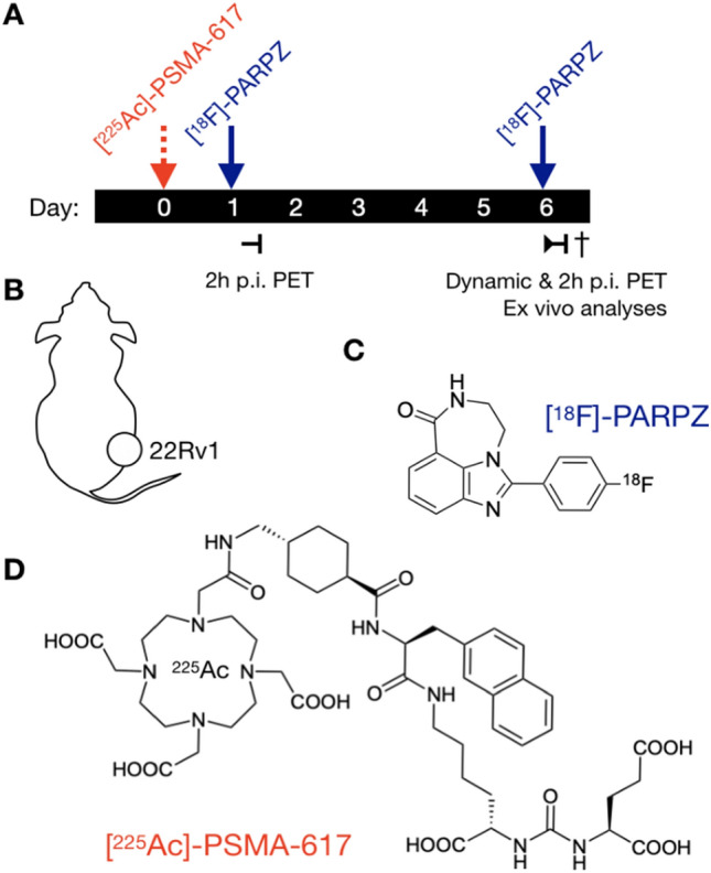
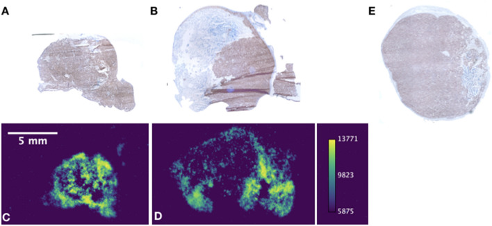
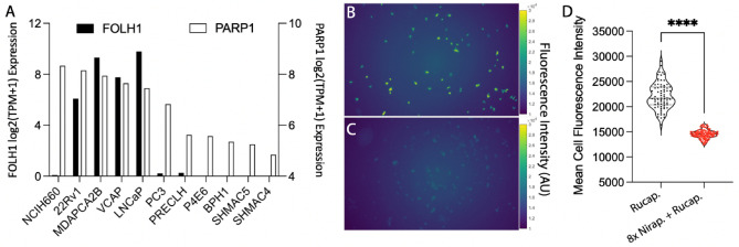
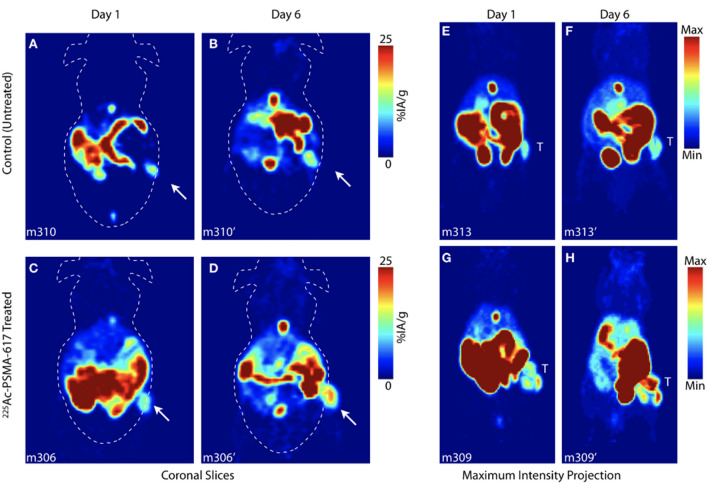
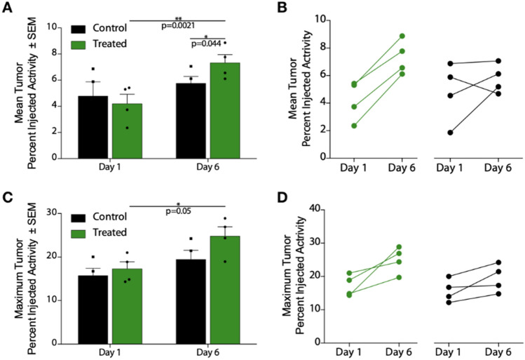
The growing interest and clinical translation of alpha particle (α) therapies brings with it new challenges to assess target cell engagement and to monitor therapeutic effect. Noninvasive imaging has great potential to guide α-treatment and to harness the potential of these agents in the complex environment of disseminated disease. Poly(ADP) ribose polymerase 1 (PARP-1) is among the most abundantly expressed DNA repair enzymes with key roles in multiple repair pathways-such as those induced by irradiation. Here, we used a third-generation PARP1-specific radiotracer, [F]-PARPZ, to delineate castrate resistant prostate cancer xenografts. Following treatment with the clinically applied [Ac]-PSMA-617, positron emission tomography was performed and correlative autoradiography and histology acquired. [F]-PARPZ was able to distinguish treated from control (saline) xenografts by increased uptake. Kinetic analysis of tracer accumulation also suggests that the localization of the agent to sites of increased PARP-1 expression is a consequence of DNA damage response. Together, these data support expanded investigation of [F]-PARPZ to facilitate clinical translation in the ⍺-therapy space.
α 粒子(α)疗法日益受到关注并逐渐走向临床应用,这为评估靶细胞结合情况和监测治疗效果带来了新的挑战。非侵入性成像技术在指导 α 治疗和挖掘这些药物在广泛疾病复杂环境中的潜力方面具有巨大的潜力。多聚(ADP-核糖)聚合酶 1(PARP-1)是表达最丰富的 DNA 修复酶之一,在多种修复途径中发挥关键作用,如辐射诱导的修复途径。在这里,我们使用了第三代 PARP1 特异性放射性示踪剂 [F]-PARPZ,来描绘去势抵抗性前列腺癌异种移植物。在用临床应用的 [Ac]-PSMA-617 治疗后,进行正电子发射断层扫描,并获得放射性自显影和组织学的相关性分析。[F]-PARPZ 通过增加摄取来区分治疗组和对照组(生理盐水)异种移植物。示踪剂积累的动力学分析也表明,该药物定位于 PARP-1 表达增加的部位是 DNA 损伤反应的结果。这些数据共同支持进一步研究 [F]-PARPZ,以促进在 α 治疗领域的临床转化。