Department of General Surgery, The Second Affiliated Hospital of Nanjing Medical University, No. 121, Jiangjiayuan Road, Nanjing, 210011, Jiangsu, China.
Dig Dis Sci. 2023 Apr;68(4):1473-1481. doi: 10.1007/s10620-022-07640-3. Epub 2022 Jul 31.
Computed tomography is the most commonly used imaging modality for preoperative assessment of lymph node status, but the reported accuracy is unsatisfactory.
To evaluate and verify the predictive performance of computed tomography deep learning on the presurgical evaluation of lymph node metastasis in patients with gastric cancer.
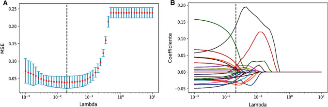
347 patients were retrospectively selected (training cohort: 242, test cohort: 105). The enhanced computed tomography arterial phase images of gastric cancer were used for lesion segmentation, radiomics and deep learning feature extraction. Three methods were used for feature selection. Support vector machine (SVM) or random forest (RF) was used to build models. The classification performance of the models was evaluated using the area under the receiver operating characteristic curve (AUC). We also established a nomogram that included clinical predictors.
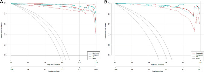
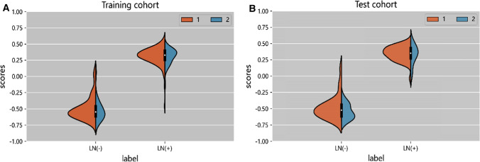
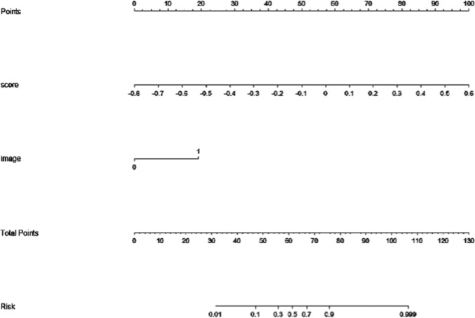
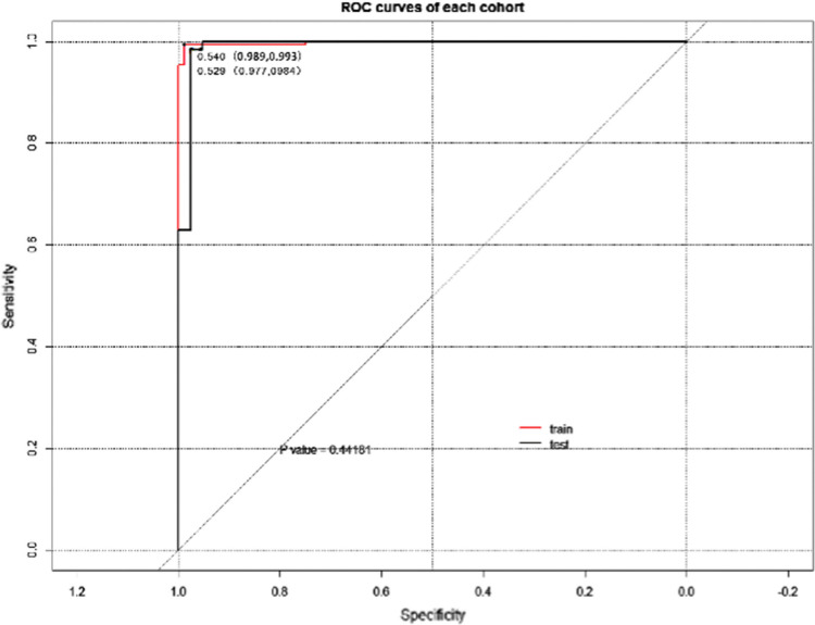
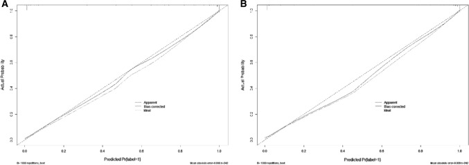
The model based on ResNet50-RF showed favorable classification performance and was verified in the test cohort (AUC = 0.9803). The nomogram based on deep learning feature scores and the lymph node status reported by computed tomography showed excellent discrimination. AUC of 0.9978 was achieved in the training cohort and verified in the test cohort (AUC = 0.9914). Decision analysis curve showed the value of nomogram in clinical application.
The computed tomography-based deep learning nomogram can accurately and effectively evaluate lymph node metastasis in patients with gastric cancer before surgery.
计算机断层扫描是术前评估淋巴结状态最常用的成像方式,但报道的准确性并不令人满意。
评估和验证计算机断层扫描深度学习在胃癌患者术前评估淋巴结转移中的预测性能。
回顾性选择了 347 名患者(训练队列:242 名,测试队列:105 名)。使用增强 CT 动脉期图像对胃癌病灶进行分割、放射组学和深度学习特征提取。使用三种方法进行特征选择。支持向量机(SVM)或随机森林(RF)用于构建模型。使用受试者工作特征曲线下的面积(AUC)评估模型的分类性能。我们还建立了一个包含临床预测因子的列线图。
基于 ResNet50-RF 的模型表现出良好的分类性能,并在测试队列中得到验证(AUC=0.9803)。基于深度学习特征评分和 CT 报告的淋巴结状态的列线图具有出色的判别能力。在训练队列中获得了 AUC 为 0.9978 的结果,并在测试队列中得到验证(AUC=0.9914)。决策分析曲线显示了列线图在临床应用中的价值。
基于 CT 的深度学习列线图可准确有效地评估胃癌患者术前的淋巴结转移情况。