• 文献检索
  • 文档翻译
  • 深度研究
  • 学术资讯
  • Suppr Zotero 插件Zotero 插件
  • 邀请有礼
  • 套餐&价格
  • 历史记录
应用&插件
Suppr Zotero 插件Zotero 插件浏览器插件Mac 客户端Windows 客户端微信小程序
定价
高级版会员购买积分包购买API积分包
服务
文献检索文档翻译深度研究API 文档MCP 服务
关于我们
关于 Suppr公司介绍联系我们用户协议隐私条款
关注我们

Suppr 超能文献

核心技术专利:CN118964589B侵权必究
粤ICP备2023148730 号-1Suppr @ 2026

文献检索

告别复杂PubMed语法,用中文像聊天一样搜索,搜遍4000万医学文献。AI智能推荐,让科研检索更轻松。

立即免费搜索

文件翻译

保留排版,准确专业,支持PDF/Word/PPT等文件格式,支持 12+语言互译。

免费翻译文档

深度研究

AI帮你快速写综述,25分钟生成高质量综述,智能提取关键信息,辅助科研写作。

立即免费体验

基于 CT 图像分割的放射组学框架的建立与验证。

Development and verification of radiomics framework for computed tomography image segmentation.

机构信息

Southeast University, Laboratory of Image Science and Technology, Jiangsu Provincial Joint International Research Laboratory of Medical Information Processing, Centre de Recherche en Information Biomédicale Sino-français (CRIBs), Nanjing, P. R. China.

Department of Radiation Oncology Physics and Technology, Shandong Cancer Hospital and Institute, Shandong First Medical University and Shandong Academy of Medical Sciences, Jinan, China.

出版信息

Med Phys. 2022 Oct;49(10):6527-6537. doi: 10.1002/mp.15904. Epub 2022 Aug 17.

DOI:10.1002/mp.15904
PMID:35917213
原文链接:https://pmc.ncbi.nlm.nih.gov/articles/PMC9805121/
Abstract

BACKGROUND

Radiomics has been considered an imaging marker for capturing quantitative image information (QII). The introduction of radiomics to image segmentation is desirable but challenging.

PURPOSE

This study aims to develop and validate a radiomics-based framework for image segmentation (RFIS).

METHODS

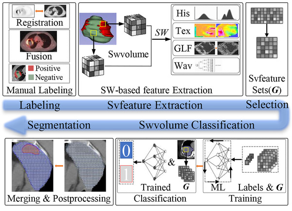
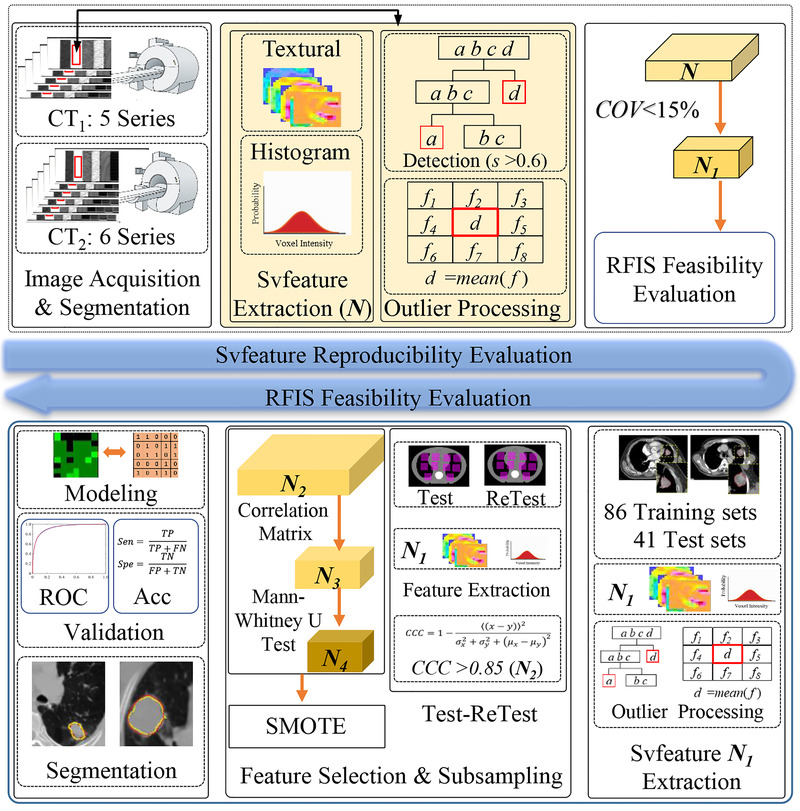
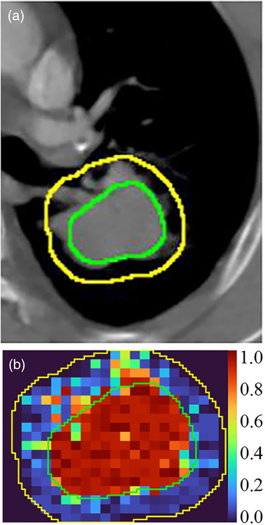
RFIS is designed using features extracted from volume (svfeatures) created by sliding window (swvolume). The 53 svfeatures are extracted from 11 phantom series. Outliers in the svfeature datasets are detected by isolation forest (iForest) and specified as the mean value. The percentage coefficient of variation (%COV) is calculated to evaluate the reproducibility of svfeatures. RFIS is constructed and applied to the gross target volume (GTV) segmentation from the peritumoral region (GTV with a 10 mm margin) to assess its feasibility. The 127 lung cancer images are enrolled. The test-retest method, correlation matrix, and Mann-Whitney U test (p < 0.05) are used to select non-redundant svfeatures of statistical significance from the reproducible svfeatures. The synthetic minority over-sampling technique is utilized to balance the minority group in the training sets. The support vector machine is employed for RFIS construction, which is tuned in the training set using 10-fold stratified cross-validation and then evaluated in the test sets. The swvolumes with the consistent classification results are grouped and merged. Mode filtering is performed to remove very small subvolumes and create relatively large regions of completely uniform character. In addition, RFIS performance is evaluated by the area under the receiver operating characteristic (ROC) curve (AUC), accuracy, sensitivity, specificity, and Dice similarity coefficient (DSC).

RESULTS

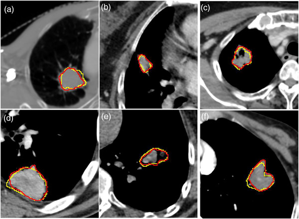
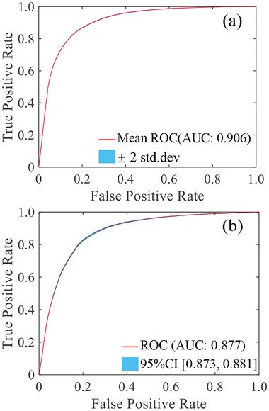
30249 phantom and 145008 patient image swvolumes were analyzed. Forty-nine (92.45% of 53) svfeatures represented excellent reproducibility(%COV<15). Forty-five features (91.84% of 49) included five categories that passed test-retest analysis. Thirteen svfeatures (28.89% of 45) svfeatures were selected for RFIS construction. RFIS showed an average (95% confidence interval) sensitivity of 0.848 (95% CI:0.844-0.883), a specificity of 0.821 (95% CI: 0.818-0.825), an accuracy of 83.48% (95% CI: 83.27%-83.70%), and an AUC of 0.906 (95% CI: 0.904-0.908) with cross-validation. The sensitivity, specificity, accuracy, and AUC were equal to 0.762 (95% CI: 0.754-0.770), 0.840 (95% CI: 0.837-0.844), 82.29% (95% CI: 81.90%-82.60%), and 0.877 (95% CI: 0.873-0.881) in the test set, respectively. GTV was segmented by grouping and merging swvolume with identical classification results. The mean DSC after mode filtering was 0.707 ± 0.093 in the training sets and 0.688 ± 0.072 in the test sets.

CONCLUSION

Reproducible svfeatures can capture the differences in QII among swvolumes. RFIS can be applied to swvolume classification, which achieves image segmentation by grouping and merging the swvolume with similar QII.

摘要

背景

放射组学被认为是一种捕获定量图像信息(QII)的成像标志物。将放射组学引入图像分割是可取的,但具有挑战性。

目的

本研究旨在开发和验证一种基于放射组学的图像分割框架(RFIS)。

方法

RFIS 是使用从滑动窗口(swvolume)创建的体积(svfeatures)中提取的特征设计的。从 11 个体模系列中提取了 53 个 svfeatures。离群值通过隔离森林(iForest)检测并指定为平均值。计算百分比变异系数(%COV)以评估 svfeatures 的可重复性。构建 RFIS 并将其应用于从肿瘤周围区域(带有 10mm 边界的 GTV)分割大体肿瘤体积(GTV),以评估其可行性。共纳入 127 例肺癌图像。使用测试-再测试方法、相关矩阵和曼-惠特尼 U 检验(p<0.05)从可重复的 svfeatures 中选择具有统计学意义的非冗余 svfeatures。利用合成少数过采样技术平衡训练集中的少数群体。支持向量机用于 RFIS 构建,在训练集中使用 10 折分层交叉验证进行调整,然后在测试集中进行评估。具有一致分类结果的 swvolume 被分组和合并。模式过滤用于去除非常小的子体积并创建具有完全一致特征的相对较大区域。此外,通过接收器操作特征(ROC)曲线下面积(AUC)、准确性、灵敏度、特异性和 Dice 相似系数(DSC)评估 RFIS 的性能。

结果

分析了 30249 个体模和 145008 个患者图像 swvolume。有 49 个(53 个中的 92.45%)svfeatures 表现出极好的可重复性(%COV<15)。有 45 个特征(49 个中的 91.84%)包括通过测试-再测试分析的五类。有 13 个 svfeatures(45 个中的 28.89%)被选为 RFIS 构建。RFIS 显示平均(95%置信区间)灵敏度为 0.848(95%CI:0.844-0.883),特异性为 0.821(95%CI:0.818-0.825),准确性为 83.48%(95%CI:0.8327%-0.8370%),AUC 为 0.906(95%CI:0.904-0.908),交叉验证。在测试集中,灵敏度、特异性、准确性和 AUC 分别等于 0.762(95%CI:0.754-0.770)、0.840(95%CI:0.837-0.844)、82.29%(95%CI:81.90%-82.60%)和 0.877(95%CI:0.873-0.881)。通过分组和合并具有相同分类结果的 swvolume 对 GTV 进行分割。模式过滤后的平均 DSC 在训练集中为 0.707±0.093,在测试集中为 0.688±0.072。

结论

可重复的 svfeatures 可以捕获 swvolume 之间 QII 的差异。RFIS 可应用于 swvolume 分类,通过分组和合并具有相似 QII 的 swvolume 来实现图像分割。

https://cdn.ncbi.nlm.nih.gov/pmc/blobs/3269/9805121/fe23ac2c6215/MP-49-6527-g001.jpg
https://cdn.ncbi.nlm.nih.gov/pmc/blobs/3269/9805121/ae351b9a705d/MP-49-6527-g003.jpg
https://cdn.ncbi.nlm.nih.gov/pmc/blobs/3269/9805121/ddf550a7b972/MP-49-6527-g005.jpg
https://cdn.ncbi.nlm.nih.gov/pmc/blobs/3269/9805121/38d3eb77283b/MP-49-6527-g002.jpg
https://cdn.ncbi.nlm.nih.gov/pmc/blobs/3269/9805121/68dba9dcf206/MP-49-6527-g004.jpg
https://cdn.ncbi.nlm.nih.gov/pmc/blobs/3269/9805121/fe23ac2c6215/MP-49-6527-g001.jpg

相似文献

1
Development and verification of radiomics framework for computed tomography image segmentation.基于 CT 图像分割的放射组学框架的建立与验证。
Med Phys. 2022 Oct;49(10):6527-6537. doi: 10.1002/mp.15904. Epub 2022 Aug 17.
2
High quality machine-robust image features: identification in nonsmall cell lung cancer computed tomography images.高质量的机器鲁棒图像特征:非小细胞肺癌 CT 图像中的识别。
Med Phys. 2013 Dec;40(12):121916. doi: 10.1118/1.4829514.
3
Reproducibility and non-redundancy of radiomic features extracted from arterial phase CT scans in hepatocellular carcinoma patients: impact of tumor segmentation variability.肝细胞癌患者动脉期CT扫描提取的影像组学特征的可重复性和非冗余性:肿瘤分割变异性的影响
Quant Imaging Med Surg. 2019 Mar;9(3):453-464. doi: 10.21037/qims.2019.03.02.
4
Radiomics as a measure superior to common similarity metrics for tumor segmentation performance evaluation.放射组学作为一种优于常用相似性度量的肿瘤分割性能评估方法。
J Appl Clin Med Phys. 2024 Aug;25(8):e14442. doi: 10.1002/acm2.14442. Epub 2024 Jun 23.
5
Multi-site quality and variability analysis of 3D FDG PET segmentations based on phantom and clinical image data.基于体模和临床图像数据的3D FDG PET分割的多中心质量与变异性分析
Med Phys. 2017 Feb;44(2):479-496. doi: 10.1002/mp.12041.
6
Computer-aided diagnosis of ground glass pulmonary nodule by fusing deep learning and radiomics features.基于深度学习和放射组学特征融合的磨玻璃肺结节计算机辅助诊断。
Phys Med Biol. 2021 Mar 4;66(6):065015. doi: 10.1088/1361-6560/abe735.
7
Computed Tomography-Based Radiomics Model to Predict Central Cervical Lymph Node Metastases in Papillary Thyroid Carcinoma: A Multicenter Study.基于计算机断层扫描的影像组学模型预测甲状腺乳头状癌中央颈部淋巴结转移:一项多中心研究。
Front Endocrinol (Lausanne). 2021 Oct 21;12:741698. doi: 10.3389/fendo.2021.741698. eCollection 2021.
8
CT-based radiomics and machine learning to predict spread through air space in lung adenocarcinoma.基于 CT 的放射组学和机器学习预测肺腺癌的空气空间播散。
Eur Radiol. 2020 Jul;30(7):4050-4057. doi: 10.1007/s00330-020-06694-z. Epub 2020 Feb 28.
9
Influence of segmentation margin on machine learning-based high-dimensional quantitative CT texture analysis: a reproducibility study on renal clear cell carcinomas.基于分割边界的机器学习高维定量 CT 纹理分析的影响:对肾透明细胞癌的可重复性研究。
Eur Radiol. 2019 Sep;29(9):4765-4775. doi: 10.1007/s00330-019-6003-8. Epub 2019 Feb 12.
10
Automatic segmentation of bladder cancer on MRI using a convolutional neural network and reproducibility of radiomics features: a two-center study.基于卷积神经网络的 MRI 膀胱癌自动分割及影像组学特征的可重复性:一项双中心研究。
Sci Rep. 2023 Jan 12;13(1):628. doi: 10.1038/s41598-023-27883-y.

本文引用的文献

1
Recent advances and clinical applications of deep learning in medical image analysis.深度学习在医学图像分析中的最新进展和临床应用。
Med Image Anal. 2022 Jul;79:102444. doi: 10.1016/j.media.2022.102444. Epub 2022 Apr 4.
2
Gross Tumor Volume Segmentation for Stage III NSCLC Radiotherapy Using 3D ResSE-Unet.使用3D ResSE-Unet进行III期非小细胞肺癌放疗的肿瘤总体积分割
Technol Cancer Res Treat. 2022 Jan-Dec;21:15330338221090847. doi: 10.1177/15330338221090847.
3
A radiomics-boosted deep-learning model for COVID-19 and non-COVID-19 pneumonia classification using chest x-ray images.
基于放射组学增强的深度学习模型,利用胸部 X 光图像对 COVID-19 和非 COVID-19 肺炎进行分类。
Med Phys. 2022 May;49(5):3213-3222. doi: 10.1002/mp.15582. Epub 2022 Mar 15.
4
Lung cancer diagnosis using deep attention-based multiple instance learning and radiomics.基于深度注意力的多实例学习和放射组学在肺癌诊断中的应用。
Med Phys. 2022 May;49(5):3134-3143. doi: 10.1002/mp.15539. Epub 2022 Mar 3.
5
Intratumoral analysis of digital breast tomosynthesis for predicting the Ki-67 level in breast cancer: A multi-center radiomics study.基于数字乳腺断层合成术的肿瘤内分析预测乳腺癌 Ki-67 水平:一项多中心放射组学研究。
Med Phys. 2022 Jan;49(1):219-230. doi: 10.1002/mp.15392. Epub 2021 Dec 13.
6
Subregional radiomics analysis for the detection of the EGFR mutation on thoracic spinal metastases from lung cancer.用于检测肺癌胸椎转移灶中表皮生长因子受体(EGFR)突变的亚区域放射组学分析
Phys Med Biol. 2021 Oct 26;66(21). doi: 10.1088/1361-6560/ac2ea7.
7
The Impact of Artificial Intelligence CNN Based Denoising on FDG PET Radiomics.基于人工智能卷积神经网络去噪对氟代脱氧葡萄糖正电子发射断层扫描影像组学的影响
Front Oncol. 2021 Aug 24;11:692973. doi: 10.3389/fonc.2021.692973. eCollection 2021.
8
Combining computed tomography and biologically effective dose in radiomics and deep learning improves prediction of tumor response to robotic lung stereotactic body radiation therapy.将计算机断层扫描与生物有效剂量相结合进行放射组学和深度学习可提高对机器人肺部立体定向体放射治疗肿瘤反应的预测。
Med Phys. 2021 Oct;48(10):6257-6269. doi: 10.1002/mp.15178. Epub 2021 Sep 2.
9
Automatic segmentation of lung tumors on CT images based on a 2D & 3D hybrid convolutional neural network.基于二维和三维混合卷积神经网络的 CT 图像肺肿瘤自动分割。
Br J Radiol. 2021 Oct 1;94(1126):20210038. doi: 10.1259/bjr.20210038. Epub 2021 Aug 4.
10
Simultaneous Identification of and Mutations in Patients with Non-Small Cell Lung Cancer by Machine Learning-Derived Three-Dimensional Radiomics.通过机器学习衍生的三维放射组学同时鉴定非小细胞肺癌患者中的 和 突变
Cancers (Basel). 2021 Apr 10;13(8):1814. doi: 10.3390/cancers13081814.