You Wei, Sun Yong, Feng Junqiang, Wang Zhiliang, Li Lin, Chen Xiheng, Lv Jian, Tang Yudi, Deng Dingwei, Wei Dachao, Gui Siming, Liu Xinke, Liu Peng, Jin Hengwei, Ge Huijian, Zhang Yanling
Department of Interventional Neuroradiology, Beijing Neurosurgical Institute and Beijing Tiantan Hospital, Capital Medical University, Beijing, China.
Department of Neurointerventional Engineering and Technology, Beijing Engineering Research Center, Beijing, China.
Front Neurol. 2022 Jul 19;13:932933. doi: 10.3389/fneur.2022.932933. eCollection 2022.
Unruptured intracranial aneurysms (UIAs) are increasingly being detected in clinical practice. Artificial intelligence (AI) has been increasingly used to assist diagnostic techniques and shows encouraging prospects. In this study, we reported the protocol and preliminary results of the establishment of an intracranial aneurysm database for AI application based on computed tomography angiography (CTA) images.
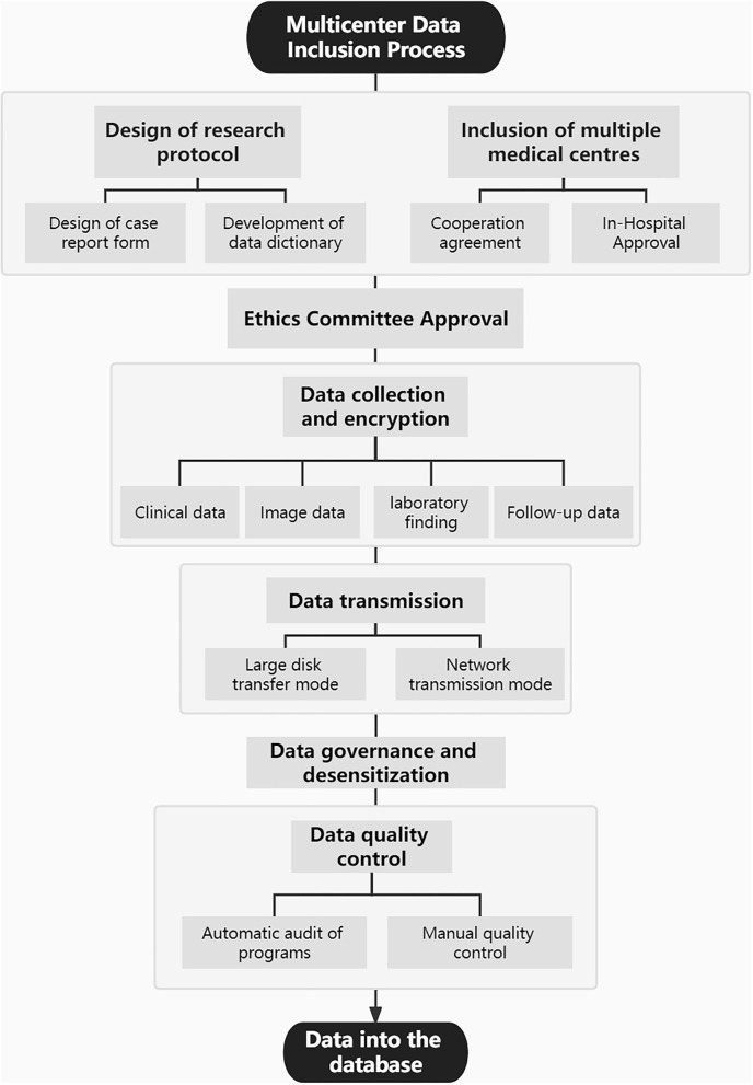
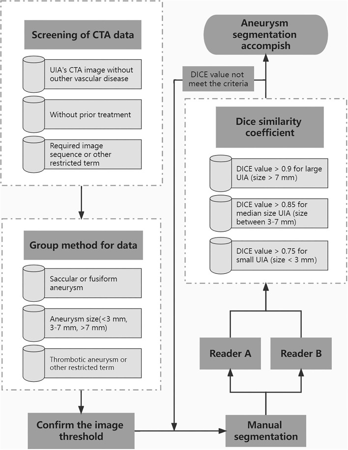
Through a review of picture archiving and communication systems, we collected CTA images of patients with aneurysms between January 2010 and March 2021. The radiologists performed manual segmentation of all diagnosed aneurysms on subtraction CTA as the basis for automatic aneurysm segmentation. Then, AI will be applied to two stages of aneurysm treatment, namely, automatic aneurysm detection and segmentation model based on the CTA image and the aneurysm risk prediction model.
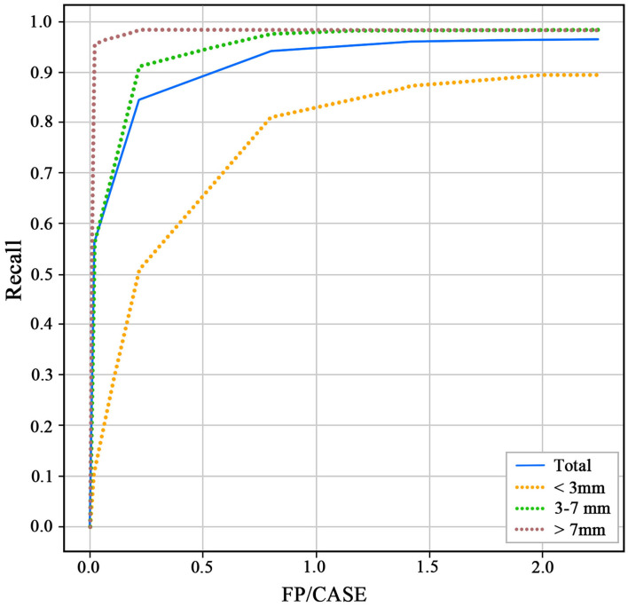
Three medical centers have been included in this study so far. A total of 3,190 cases of CTA examinations with 4,124 aneurysms were included in the database. All identified aneurysms from CTA images that enrolled in this study were manually segmented on subtraction CTA by six readers. We developed a structure of 3D-Unet for aneurysm detection and segmentation in CTA images. The algorithm was developed and tested using a total of 2,272 head CTAs with 2,938 intracranial aneurysms. The recall and false positives per case (FP/case) of this model for detecting aneurysms were 0.964 and 2.01, and the Dice values for aneurysm segmentation were 0.783.
This study introduces the protocol and preliminary results of the establishment of the intracranial aneurysm database for AI applications based on CTA images. The establishment of a multicenter database based on CTA images of intracranial aneurysms is the basis for the application of AI in the diagnosis and treatment of aneurysms. In addition to segmentation, AI should have great potential for aneurysm treatment and management in the future.
未破裂颅内动脉瘤(UIAs)在临床实践中越来越多地被检测出来。人工智能(AI)已越来越多地用于辅助诊断技术,并显示出令人鼓舞的前景。在本研究中,我们报告了基于计算机断层扫描血管造影(CTA)图像建立用于AI应用的颅内动脉瘤数据库的方案和初步结果。
通过回顾图像存档与通信系统,我们收集了2010年1月至2021年3月期间动脉瘤患者的CTA图像。放射科医生在减影CTA上对所有诊断出的动脉瘤进行手动分割,作为自动动脉瘤分割的基础。然后,AI将应用于动脉瘤治疗的两个阶段,即基于CTA图像的自动动脉瘤检测和分割模型以及动脉瘤风险预测模型。
到目前为止,本研究已纳入三个医学中心。数据库中总共包括3190例CTA检查,其中有4124个动脉瘤。参与本研究的CTA图像中所有识别出的动脉瘤均由六位阅片者在减影CTA上进行手动分割。我们开发了一种用于CTA图像中动脉瘤检测和分割的3D-Unet结构。该算法使用总共2272例头部CTA和2938个颅内动脉瘤进行开发和测试。该模型检测动脉瘤的召回率和每例假阳性(FP/例)分别为0.964和2.01,动脉瘤分割的Dice值为0.783。
本研究介绍了基于CTA图像建立用于AI应用的颅内动脉瘤数据库的方案和初步结果。基于颅内动脉瘤CTA图像建立多中心数据库是AI在动脉瘤诊断和治疗中应用的基础。除分割外,AI在未来动脉瘤治疗和管理中应具有巨大潜力。