Nakamura Masahiko, Murasato Futa, Øverby Anders, Kodama Yosuke, Michimae Hirofumi, Sasaki Kazuki, Flahou Bram, Haesebrouck Freddy, Murayama Somay Y, Takahashi Shinichi, Uchida Masayuki, Suzuki Hidekazu, Matsui Hidenori
Ohmura Satoshi Memorial Institute, Kitasato University, Tokyo, Japan.
School of Pharmacy, Kitasato University, Tokyo, Japan.
Front Pharmacol. 2022 Jul 22;13:692437. doi: 10.3389/fphar.2022.692437. eCollection 2022.
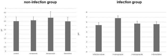
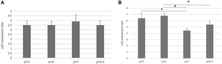
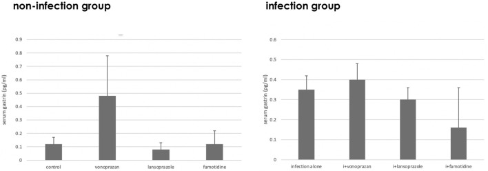
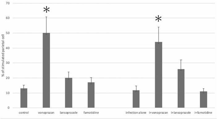
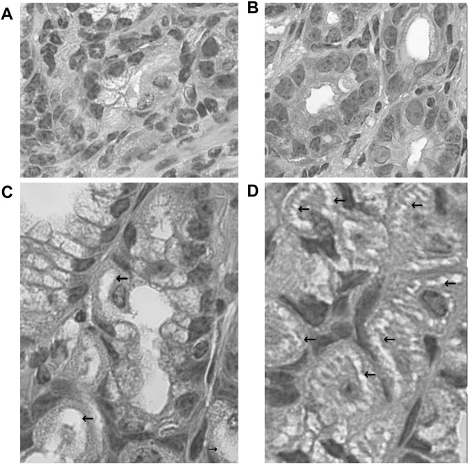
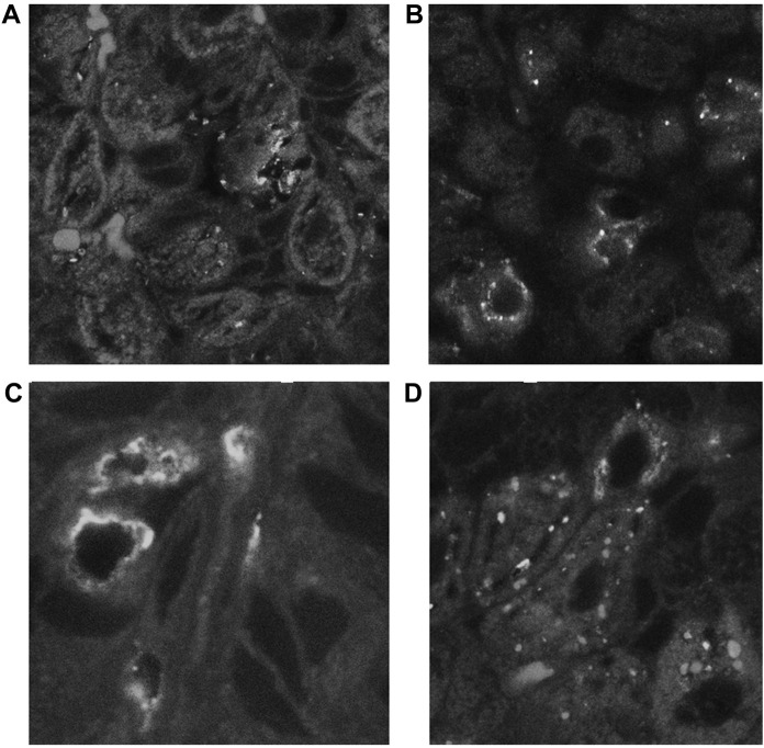
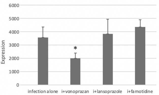
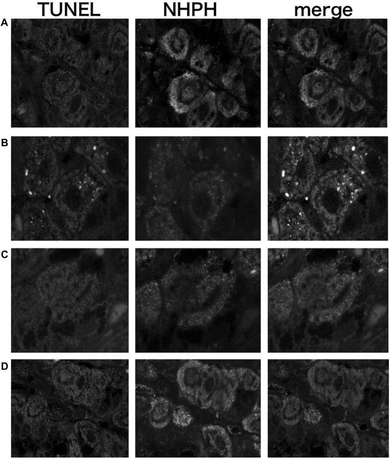
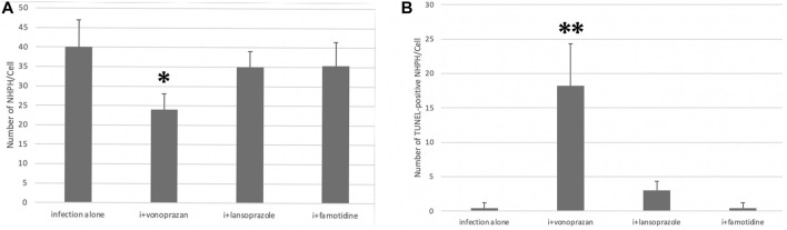
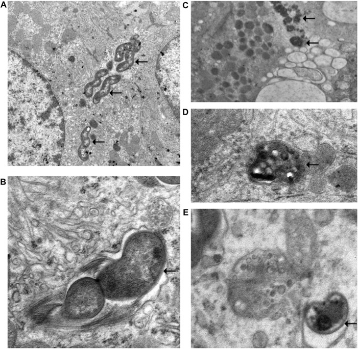
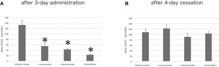
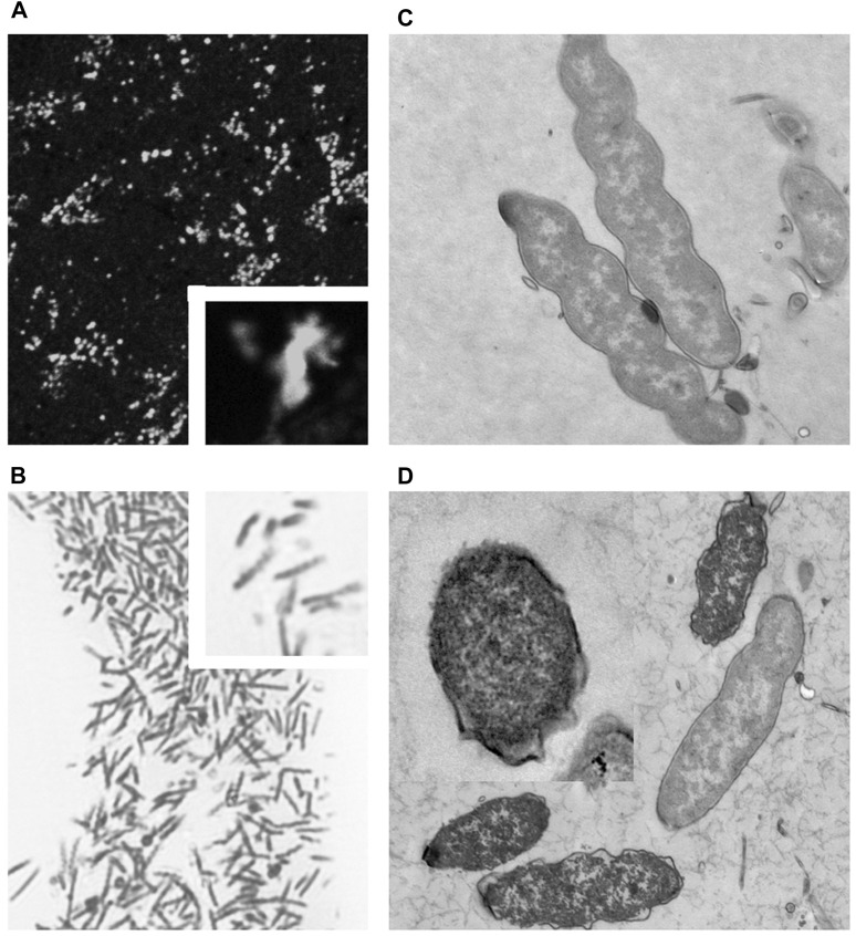
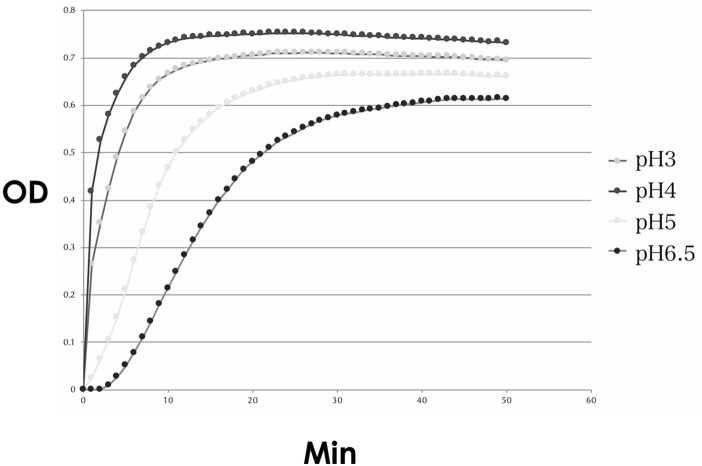
We investigated the effect of increased pH induced by acid suppressants on the viability of non helicobacters (NHPHs) within parietal cell intracellular canaliculi and fundic glandular lumina by immunohistochemistry, electron microscopy, quantitative PCR, urea breath tests, and using a bilayer culture system. Three months before the experiment, mice were infected with the NHPH and then treated with famotidine (2 mg/kg body weight [BW], once daily), lansoprazole (30 mg/kg BW, once daily), or vonoprazan (20 mg/kg BW, once daily) for 3 days. Immunohistochemical studies using the TUNEL method, quantitative PCR analysis, and urea breath tests were performed. PCR analysis showed a decrease in the NHPH quantity after vonoprazan treatment. Urea breath tests revealed a significant decrease in the NHPH urease activity after vonoprazan, lansoprazole, and famotidine treatments for 3 days; however, 4 days after the treatment, urease activity reversed to the pretreatment level for each treatment group. Electron microscopy revealed an increase in the damaged NHPH after vonoprazan treatment. The TUNEL method revealed apoptotic NHPH within parietal cells after vonoprazan treatment. The bilayer culture results demonstrated that NHPH moved more quickly at a pH of 4.0 than at a pH of 3.0, 5.0, and 6.5, and electron microscopy revealed a change from the spiral form to the coccoid form under near-neutral pH conditions. We thus proposed that acid suppressants, especially vonoprazan, induce NHPH damage by altering pH.
我们通过免疫组织化学、电子显微镜、定量聚合酶链反应(PCR)、尿素呼气试验以及使用双层培养系统,研究了抑酸剂引起的pH值升高对壁细胞内小管和胃底腺管腔中非幽门螺杆菌(NHPHs)生存能力的影响。实验前三个月,将小鼠感染NHPH,然后分别用法莫替丁(2毫克/千克体重[BW],每日一次)、兰索拉唑(30毫克/千克BW,每日一次)或沃克帕唑(20毫克/千克BW,每日一次)治疗3天。进行了使用TUNEL法的免疫组织化学研究、定量PCR分析和尿素呼气试验。PCR分析显示沃克帕唑治疗后NHPH数量减少。尿素呼气试验显示,沃克帕唑、兰索拉唑和法莫替丁治疗3天后,NHPH脲酶活性显著降低;然而,治疗4天后,各治疗组的脲酶活性恢复到治疗前水平。电子显微镜显示沃克帕唑治疗后受损的NHPH增加。TUNEL法显示沃克帕唑治疗后壁细胞内有凋亡的NHPH。双层培养结果表明,NHPH在pH值为4.0时比在pH值为3.0、5.0和6.5时移动得更快,电子显微镜显示在近中性pH条件下,NHPH从螺旋形变为球菌形。因此,我们提出抑酸剂,尤其是沃克帕唑,通过改变pH值诱导NHPH损伤。