Department of Biochemistry and Tissue Biology, Institute of Biology, University of Campinas, Campinas, 13083862, Brazil.
Ribeirão Preto Medical School, University of São Paulo, Ribeirão Preto, 14049900, Brazil.
Proc Natl Acad Sci U S A. 2022 Aug 30;119(35):e2200960119. doi: 10.1073/pnas.2200960119. Epub 2022 Aug 11.
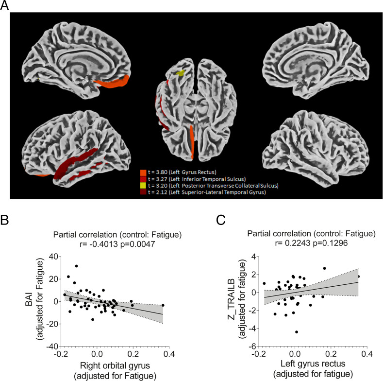
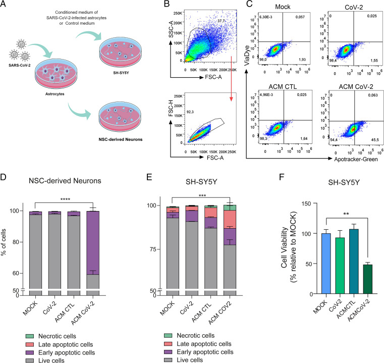
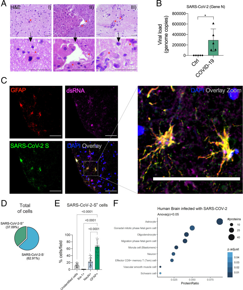
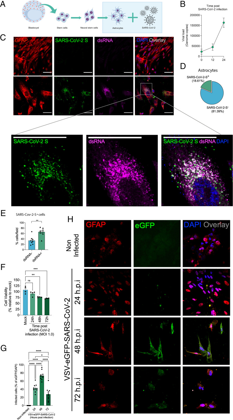
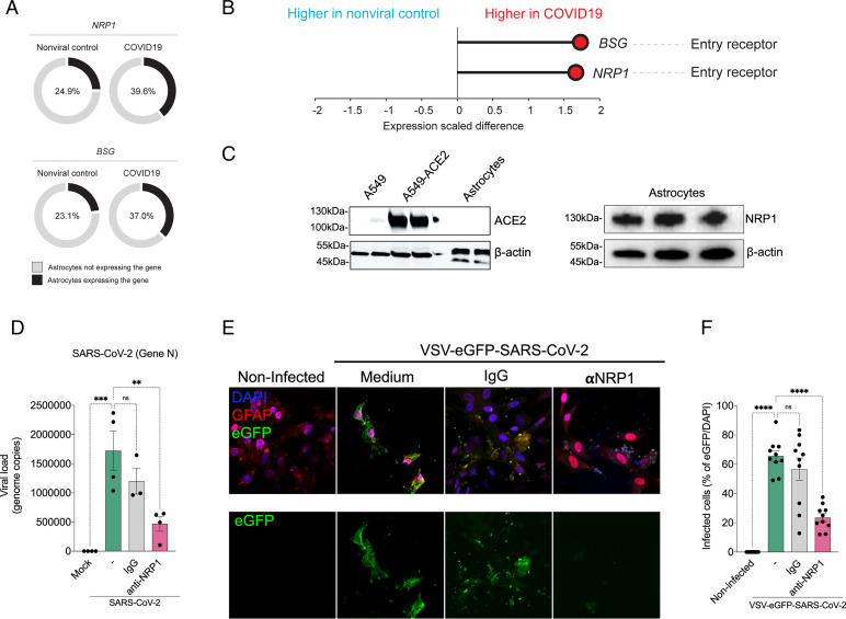
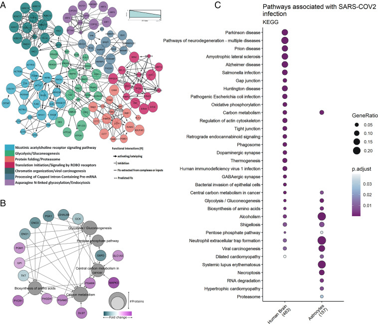
Although increasing evidence confirms neuropsychiatric manifestations associated mainly with severe COVID-19 infection, long-term neuropsychiatric dysfunction (recently characterized as part of "long COVID-19" syndrome) has been frequently observed after mild infection. We show the spectrum of cerebral impact of severe acute respiratory syndrome coronavirus 2 (SARS-CoV-2) infection, ranging from long-term alterations in mildly infected individuals (orbitofrontal cortical atrophy, neurocognitive impairment, excessive fatigue and anxiety symptoms) to severe acute damage confirmed in brain tissue samples extracted from the orbitofrontal region (via endonasal transethmoidal access) from individuals who died of COVID-19. In an independent cohort of 26 individuals who died of COVID-19, we used histopathological signs of brain damage as a guide for possible SARS-CoV-2 brain infection and found that among the 5 individuals who exhibited those signs, all of them had genetic material of the virus in the brain. Brain tissue samples from these five patients also exhibited foci of SARS-CoV-2 infection and replication, particularly in astrocytes. Supporting the hypothesis of astrocyte infection, neural stem cell-derived human astrocytes in vitro are susceptible to SARS-CoV-2 infection through a noncanonical mechanism that involves spike-NRP1 interaction. SARS-CoV-2-infected astrocytes manifested changes in energy metabolism and in key proteins and metabolites used to fuel neurons, as well as in the biogenesis of neurotransmitters. Moreover, human astrocyte infection elicits a secretory phenotype that reduces neuronal viability. Our data support the model in which SARS-CoV-2 reaches the brain, infects astrocytes, and consequently, leads to neuronal death or dysfunction. These deregulated processes could contribute to the structural and functional alterations seen in the brains of COVID-19 patients.
虽然越来越多的证据证实了与严重 COVID-19 感染相关的神经精神表现,但在轻度感染后,长期的神经精神功能障碍(最近被描述为“长 COVID-19”综合征的一部分)经常被观察到。我们展示了严重急性呼吸综合征冠状病毒 2(SARS-CoV-2)感染对大脑的影响谱,从轻度感染个体的长期改变(眶额皮质萎缩、神经认知障碍、过度疲劳和焦虑症状)到从死于 COVID-19 的个体的眶额区域(通过经鼻蝶窦入路)提取的脑组织样本中证实的严重急性损伤。在一个由 26 名死于 COVID-19 的个体组成的独立队列中,我们使用脑组织损伤的组织病理学标志作为 SARS-CoV-2 脑感染的可能指南,并发现在表现出这些迹象的 5 名个体中,所有个体的大脑中都存在病毒的遗传物质。来自这五名患者的脑组织样本也显示出 SARS-CoV-2 感染和复制的焦点,特别是在星形胶质细胞中。支持星形胶质细胞感染的假说,体外源自神经干细胞的人星形胶质细胞通过涉及刺突-NRP1 相互作用的非典型机制易感染 SARS-CoV-2。感染 SARS-CoV-2 的星形胶质细胞表现出能量代谢以及用于为神经元供能的关键蛋白质和代谢物的变化,以及神经递质的生物发生变化。此外,人星形胶质细胞感染会引发一种分泌表型,降低神经元活力。我们的数据支持 SARS-CoV-2 到达大脑、感染星形胶质细胞并由此导致神经元死亡或功能障碍的模型。这些失调的过程可能导致 COVID-19 患者大脑中观察到的结构和功能改变。