Department of Chemical Engineering, Auburn University, Auburn, Alabama 36849, United States.
College of Pharmacy, Department of Pharmaceutical Sciences, Nova Southeastern University, Lauderdale, Florida 33314, United States.
ACS Biomater Sci Eng. 2022 Sep 12;8(9):3831-3841. doi: 10.1021/acsbiomaterials.2c00285. Epub 2022 Aug 15.
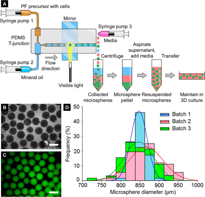
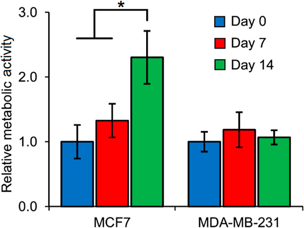
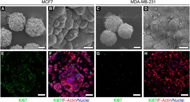
Spheroidal cancer microtissues are highly advantageous for a wide range of biomedical applications, including high-throughput drug screening, multiplexed target validation, mechanistic investigation of tumor-extracellular matrix (ECM) interactions, among others. Current techniques for spheroidal tissue formation rely heavily on self-aggregation of single cancer cells and have substantial limitations in terms of cell-type-specific heterogeneities, uniformity, ease of production and handling, and most importantly, mimicking the complex native tumor microenvironmental conditions in simplistic models. These constraints can be overcome by using engineered tunable hydrogels that closely mimic the tumor ECM and elucidate pathologically relevant cell behavior, coupled with microfluidics-based high-throughput fabrication technologies to encapsulate cells and create cancer microtissues. In this study, we employ biosynthetic hybrid hydrogels composed of poly(ethylene glycol diacrylate) (PEGDA) covalently conjugated to natural protein (fibrinogen) (PEG-fibrinogen, PF) to create monodisperse microspheres encapsulating breast cancer cells for 3D culture and tumorigenic characterization. A previously developed droplet-based microfluidic system is used for rapid, facile, and reproducible fabrication of uniform cancer microspheres with either MCF7 or MDA-MB-231 (metastatic) breast cancer cells. Cancer cell-type-dependent variations in cell viability, metabolic activity, and 3D morphology, as well as microsphere stiffness, are quantified over time. Particularly, MCF7 cells grew as tight cellular clusters in the PF microspheres, characteristic of their epithelial morphology, while MDA-MB-231 cells displayed elongated and invasive morphology, characteristic of their mesenchymal and metastatic nature. Finally, the translational potential of the cancer microsphere platform toward high-throughput drug screening is also demonstrated. With high uniformity, scalability, and control over engineered microenvironments, the established cancer microsphere model can be potentially used for mechanistic studies, fabrication of modular cancer microtissues, and future drug-testing applications.
球形癌细胞微组织在广泛的生物医学应用中具有很大的优势,包括高通量药物筛选、多重靶标验证、肿瘤细胞外基质(ECM)相互作用的机制研究等。目前用于球形组织形成的技术主要依赖于单个癌细胞的自聚集,并且在细胞类型特异性异质性、均匀性、生产和处理的便利性以及最重要的是,在简单模型中模拟复杂的天然肿瘤微环境条件方面存在很大的局限性。这些限制可以通过使用工程可调谐水凝胶来克服,这些水凝胶可以紧密模拟肿瘤 ECM,并阐明与病理相关的细胞行为,同时结合基于微流控的高通量制造技术来封装细胞并创建癌细胞微组织。在这项研究中,我们使用生物合成的杂化水凝胶,该水凝胶由共价连接天然蛋白(纤维蛋白原)的聚乙二醇二丙烯酸酯(PEGDA)组成(PEG-纤维蛋白原,PF),用于创建封装乳腺癌细胞的单分散微球,用于 3D 培养和致瘤表征。先前开发的基于液滴的微流控系统用于快速、简便和可重复地制造均匀的癌症微球,这些微球中含有 MCF7 或 MDA-MB-231(转移性)乳腺癌细胞。随着时间的推移,定量评估了细胞活力、代谢活性和 3D 形态以及微球硬度的癌症细胞类型依赖性变化。特别是,MCF7 细胞在 PF 微球中紧密聚集形成细胞簇,这是其上皮形态的特征,而 MDA-MB-231 细胞表现出伸长和侵袭形态,这是其间充质和转移性的特征。最后,还证明了癌症微球平台在高通量药物筛选中的转化潜力。该建立的癌症微球模型具有高度的均匀性、可扩展性和对工程微环境的控制,具有潜在的用于机制研究、模块化癌症微组织的制造以及未来药物测试应用的潜力。