• 文献检索
  • 文档翻译
  • 深度研究
  • 学术资讯
  • Suppr Zotero 插件Zotero 插件
  • 邀请有礼
  • 套餐&价格
  • 历史记录
应用&插件
Suppr Zotero 插件Zotero 插件浏览器插件Mac 客户端Windows 客户端微信小程序
定价
高级版会员购买积分包购买API积分包
服务
文献检索文档翻译深度研究API 文档MCP 服务
关于我们
关于 Suppr公司介绍联系我们用户协议隐私条款
关注我们

Suppr 超能文献

核心技术专利:CN118964589B侵权必究
粤ICP备2023148730 号-1Suppr @ 2026

文献检索

告别复杂PubMed语法,用中文像聊天一样搜索,搜遍4000万医学文献。AI智能推荐,让科研检索更轻松。

立即免费搜索

文件翻译

保留排版,准确专业,支持PDF/Word/PPT等文件格式,支持 12+语言互译。

免费翻译文档

深度研究

AI帮你快速写综述,25分钟生成高质量综述,智能提取关键信息,辅助科研写作。

立即免费体验

基于深度学习的术后癫痫 MRI 切除腔自动分割。

Deep learning-based automated segmentation of resection cavities on postsurgical epilepsy MRI.

机构信息

Department of Bioengineering, School of Engineering & Applied Science, University of Pennsylvania, Philadelphia, PA 19104, USA; Center for Neuroengineering and Therapeutics, University of Pennsylvania, Philadelphia, PA 19104, USA.

Center for Neuroengineering and Therapeutics, University of Pennsylvania, Philadelphia, PA 19104, USA; Department of Computer Science, University of Pennsylvania, Philadelphia, PA 19104, USA.

出版信息

Neuroimage Clin. 2022;36:103154. doi: 10.1016/j.nicl.2022.103154. Epub 2022 Aug 17.

DOI:10.1016/j.nicl.2022.103154
PMID:
35988342
原文链接:https://pmc.ncbi.nlm.nih.gov/articles/PMC9402390/
Abstract

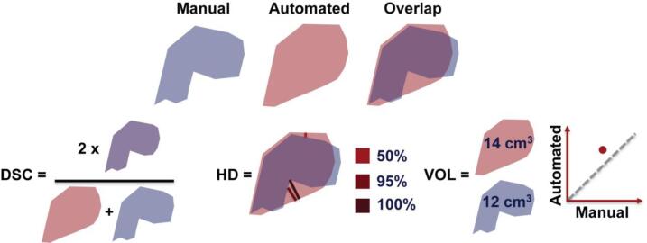
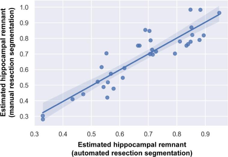
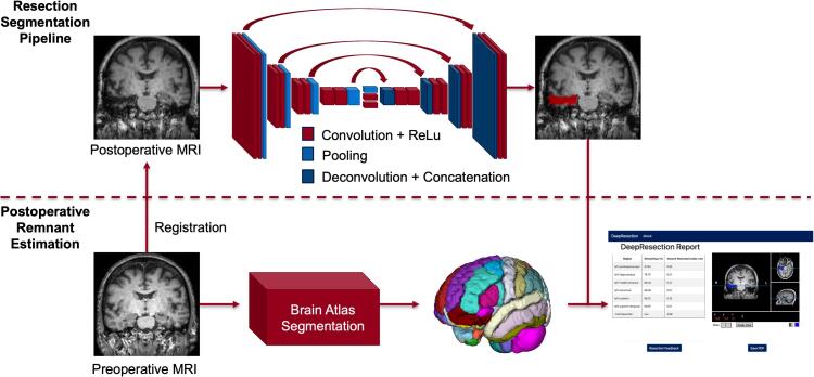
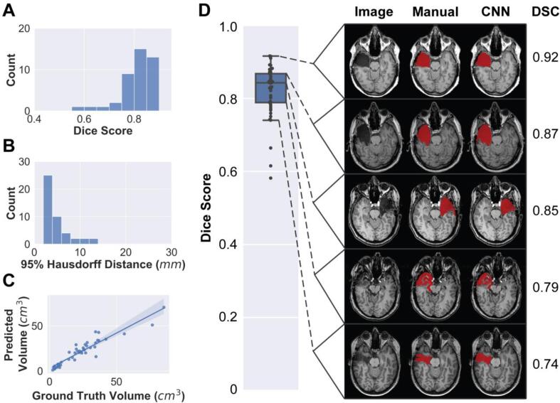
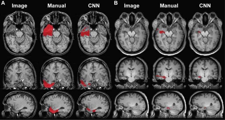
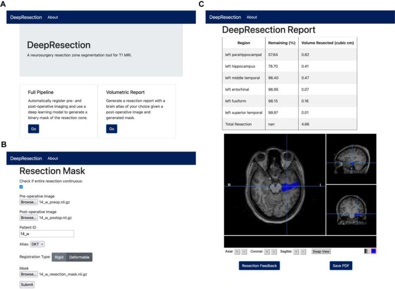
Accurate segmentation of surgical resection sites is critical for clinical assessments and neuroimaging research applications, including resection extent determination, predictive modeling of surgery outcome, and masking image processing near resection sites. In this study, an automated resection cavity segmentation algorithm is developed for analyzing postoperative MRI of epilepsy patients and deployed in an easy-to-use graphical user interface (GUI) that estimates remnant brain volumes, including postsurgical hippocampal remnant tissue. This retrospective study included postoperative T1-weighted MRI from 62 temporal lobe epilepsy (TLE) patients who underwent resective surgery. The resection site was manually segmented and reviewed by a neuroradiologist (JMS). A majority vote ensemble algorithm was used to segment surgical resections, using 3 U-Net convolutional neural networks trained on axial, coronal, and sagittal slices, respectively. The algorithm was trained using 5-fold cross validation, with data partitioned into training (N = 27) testing (N = 9), and validation (N = 9) sets, and evaluated on a separate held-out test set (N = 17). Algorithm performance was assessed using Dice-Sørensen coefficient (DSC), Hausdorff distance, and volume estimates. Additionally, we deploy a fully-automated, GUI-based pipeline that compares resection segmentations with preoperative imaging and reports estimates of resected brain structures. The cross-validation and held-out test median DSCs were 0.84 ± 0.08 and 0.74 ± 0.22 (median ± interquartile range) respectively, which approach inter-rater reliability between radiologists (0.84-0.86) as reported in the literature. Median 95 % Hausdorff distances were 3.6 mm and 4.0 mm respectively, indicating high segmentation boundary confidence. Automated and manual resection volume estimates were highly correlated for both cross-validation (r = 0.94, p < 0.0001) and held-out test subjects (r = 0.87, p < 0.0001). Automated and manual segmentations overlapped in all 62 subjects, indicating a low false negative rate. In control subjects (N = 40), the classifier segmented no voxels (N = 33), <50 voxels (N = 5), or a small volumes<0.5 cm (N = 2), indicating a low false positive rate that can be controlled via thresholding. There was strong agreement between postoperative hippocampal remnant volumes determined using automated and manual resection segmentations (r = 0.90, p < 0.0001, mean absolute error = 6.3 %), indicating that automated resection segmentations can permit quantification of postoperative brain volumes after epilepsy surgery. Applications include quantification of postoperative remnant brain volumes, correction of deformable registration, and localization of removed brain regions for network modeling.

摘要

准确的手术切除部位分割对于临床评估和神经影像学研究应用至关重要,包括切除范围的确定、手术结果的预测建模以及切除部位附近的图像处理掩模。在这项研究中,开发了一种用于分析癫痫患者术后磁共振成像的自动切除腔分割算法,并将其部署在一个易于使用的图形用户界面(GUI)中,该界面估计残余脑容量,包括术后海马残余组织。这项回顾性研究包括 62 例接受切除术的颞叶癫痫(TLE)患者的术后 T1 加权磁共振成像。切除部位由神经放射科医生(JMS)手动分割并进行评估。使用分别在轴位、冠状位和矢状位切片上训练的 3 个 U-Net 卷积神经网络,使用多数投票集成算法对手术切除部位进行分割。该算法使用 5 折交叉验证进行训练,将数据分为训练集(N=27)、测试集(N=9)和验证集(N=9),并在单独的测试集(N=17)上进行评估。使用 Dice-Sørensen 系数(DSC)、Hausdorff 距离和体积估计来评估算法性能。此外,我们还部署了一个完全自动化的基于 GUI 的管道,该管道将切除分割与术前成像进行比较,并报告切除脑结构的估计。交叉验证和测试集的中位数 DSC 分别为 0.84±0.08 和 0.74±0.22(中位数±四分位距),接近文献中报道的放射科医生之间的可靠性(0.84-0.86)。中位数 95%Hausdorff 距离分别为 3.6 毫米和 4.0 毫米,表明分割边界置信度高。交叉验证(r=0.94,p<0.0001)和测试集受试者(r=0.87,p<0.0001)的自动和手动切除体积估计高度相关。在所有 62 名受试者中,自动和手动分割都完全重叠,表明假阴性率较低。在对照组(N=40)中,分类器未分割出任何体素(N=33)、<50 个体素(N=5)或<0.5cm3 的小体积(N=2),表明假阳性率较低,可以通过阈值控制。使用自动和手动切除分割确定的术后海马残余体积之间存在很强的一致性(r=0.90,p<0.0001,平均绝对误差=6.3%),表明自动切除分割可用于量化癫痫手术后的术后残余脑容量。应用包括术后残余脑容量的量化、可变形配准的校正以及去除脑区的定位用于网络建模。

https://cdn.ncbi.nlm.nih.gov/pmc/blobs/d06f/9402390/d7c43889ebb7/gr7.jpg
https://cdn.ncbi.nlm.nih.gov/pmc/blobs/d06f/9402390/043d97c0f407/gr1.jpg
https://cdn.ncbi.nlm.nih.gov/pmc/blobs/d06f/9402390/3a34cd151ba3/gr2.jpg
https://cdn.ncbi.nlm.nih.gov/pmc/blobs/d06f/9402390/f7d092e4dadb/gr3.jpg
https://cdn.ncbi.nlm.nih.gov/pmc/blobs/d06f/9402390/08125ec47316/gr4.jpg
https://cdn.ncbi.nlm.nih.gov/pmc/blobs/d06f/9402390/ed17eb0cf2bf/gr5.jpg
https://cdn.ncbi.nlm.nih.gov/pmc/blobs/d06f/9402390/98523b1d8dc0/gr6.jpg
https://cdn.ncbi.nlm.nih.gov/pmc/blobs/d06f/9402390/d7c43889ebb7/gr7.jpg

相似文献

1
Deep learning-based automated segmentation of resection cavities on postsurgical epilepsy MRI.基于深度学习的术后癫痫 MRI 切除腔自动分割。
Neuroimage Clin. 2022;36:103154. doi: 10.1016/j.nicl.2022.103154. Epub 2022 Aug 17.
2
Automated segmentation of epilepsy surgical resection cavities: Comparison of four methods to manual segmentation.自动分割癫痫手术切除腔:四种方法与手动分割的比较。
Neuroimage. 2024 Aug 1;296:120682. doi: 10.1016/j.neuroimage.2024.120682. Epub 2024 Jun 10.
3
Multi-atlas segmentation of the whole hippocampus and subfields using multiple automatically generated templates.使用多个自动生成的模板对整个海马体及其子区进行多图谱分割。
Neuroimage. 2014 Nov 1;101:494-512. doi: 10.1016/j.neuroimage.2014.04.054. Epub 2014 Apr 29.
4
Visual ensemble selection of deep convolutional neural networks for 3D segmentation of breast tumors on dynamic contrast enhanced MRI.基于动态对比增强 MRI 的乳腺肿瘤三维分割的深度卷积神经网络的可视化集成选择。
Eur Radiol. 2023 Feb;33(2):959-969. doi: 10.1007/s00330-022-09113-7. Epub 2022 Sep 8.
5
Fully Automated MRI Segmentation and Volumetric Measurement of Intracranial Meningioma Using Deep Learning.基于深度学习的颅内脑膜瘤全自动 MRI 分割与容量测量。
J Magn Reson Imaging. 2023 Mar;57(3):871-881. doi: 10.1002/jmri.28332. Epub 2022 Jul 1.
6
Deep learning-based ultrasound auto-segmentation of the prostate with brachytherapy implanted needles.基于深度学习的放射性治疗植入针前列腺超声自动分割。
Med Phys. 2024 Apr;51(4):2665-2677. doi: 10.1002/mp.16811. Epub 2023 Oct 27.
7
Automated volumetric assessment of pituitary adenoma.垂体腺瘤的自动容积评估。
Endocrine. 2024 Jan;83(1):171-177. doi: 10.1007/s12020-023-03529-x. Epub 2023 Sep 25.
8
Using deep learning to segment breast and fibroglandular tissue in MRI volumes.利用深度学习对磁共振成像(MRI)容积中的乳腺和纤维腺组织进行分割。
Med Phys. 2017 Feb;44(2):533-546. doi: 10.1002/mp.12079.
9
Convolutional Neural Network for Automated FLAIR Lesion Segmentation on Clinical Brain MR Imaging.基于卷积神经网络的临床脑部磁共振成像 FLAIR 病变分割自动化。
AJNR Am J Neuroradiol. 2019 Aug;40(8):1282-1290. doi: 10.3174/ajnr.A6138. Epub 2019 Jul 25.
10
Clinical validation of automated hippocampal segmentation in temporal lobe epilepsy.自动海马分割在颞叶癫痫中的临床验证。
Neuroimage Clin. 2018;20:1139-1147. doi: 10.1016/j.nicl.2018.09.032. Epub 2018 Oct 10.

引用本文的文献

1
Automated generation of epilepsy surgery resection masks: The RAMPS pipeline.癫痫手术切除掩码的自动生成:RAMPS流程
Imaging Neurosci (Camb). 2025 Sep 10;3. doi: 10.1162/IMAG.a.147. eCollection 2025.
2
From Bedside to Desktop: A Data Protocol for Normative Intracranial EEG and Abnormality Mapping.从床边到桌面:一种用于规范性颅内脑电图和异常图谱的数据协议。
Bio Protoc. 2025 May 20;15(10):e5321. doi: 10.21769/BioProtoc.5321.
3
Brain Volume Loss After Stereotactic Laser Interstitial Thermal Therapy in Patients With Temporal Lobe Epilepsy.
颞叶癫痫患者立体定向激光间质热疗后的脑容量损失
J Neuroimaging. 2025 Mar-Apr;35(2):e70039. doi: 10.1111/jon.70039.
4
Swarm learning with weak supervision enables automatic breast cancer detection in magnetic resonance imaging.基于弱监督的群体学习实现磁共振成像中乳腺癌的自动检测。
Commun Med (Lond). 2025 Feb 6;5(1):38. doi: 10.1038/s43856-024-00722-5.
5
The impact of resective epilepsy surgery on the brain network: evidence from post-surgical imaging.切除性癫痫手术对脑网络的影响:来自术后影像学的证据。
Brain. 2025 Jun 3;148(6):1866-1875. doi: 10.1093/brain/awaf026.
6
Artificial intelligence in epilepsy - applications and pathways to the clinic.人工智能在癫痫中的应用及向临床应用的转化。
Nat Rev Neurol. 2024 Jun;20(6):319-336. doi: 10.1038/s41582-024-00965-9. Epub 2024 May 8.
7
Fully automatic segmentation of brain lacunas resulting from resective surgery using a 3D deep learning model.使用3D深度学习模型对切除术后脑腔隙进行全自动分割。
medRxiv. 2024 Apr 17:2023.11.16.23298572. doi: 10.1101/2023.11.16.23298572.
8
Multi-shell connectome DWI-based graph theory measures for the prediction of temporal lobe epilepsy and cognition.多壳连接体 DWI 基图论测量用于预测颞叶癫痫和认知功能。
Cereb Cortex. 2023 Jun 8;33(12):8056-8065. doi: 10.1093/cercor/bhad098.