Bogunović Hrvoje, Mares Virginia, Reiter Gregor S, Schmidt-Erfurth Ursula
Laboratory for Ophthalmic Image Analysis, Department of Ophthalmology, Medical University of Vienna, Vienna, Austria.
Department of Ophthalmology, Federal University of Minas Gerais, Belo Horizonte, Brazil.
Front Med (Lausanne). 2022 Aug 9;9:958469. doi: 10.3389/fmed.2022.958469. eCollection 2022.
To predict visual outcomes and treatment needs in a treat & extend (T&E) regimen in neovascular age-related macular degeneration (nAMD) using a machine learning model based on quantitative optical coherence tomography (OCT) imaging biomarkers.
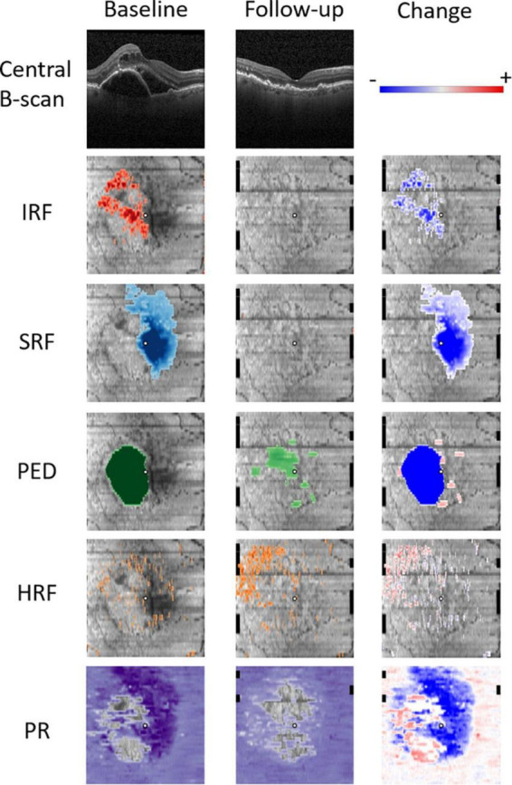
Study eyes of 270 treatment-naïve subjects, randomized to receiving ranibizumab therapy in the T&E arm of a randomized clinical trial were considered. OCT volume scans were processed at baseline and at the first follow-up visit 4 weeks later. Automated image segmentation was performed, where intraretinal (IRF), subretinal (SRF) fluid, pigment epithelial detachment (PED), hyperreflective foci, and the photoreceptor layer were delineated using a convolutional neural network (CNN). A set of respective quantitative imaging biomarkers were computed across an Early Treatment Diabetic Retinopathy Study (ETDRS) grid to describe the retinal pathomorphology spatially and its change after the first injection. Lastly, using the computed set of OCT features and available clinical and demographic information, predictive models of outcomes and retreatment intervals were built using machine learning and their performance evaluated with a 10-fold cross-validation.
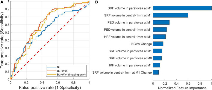
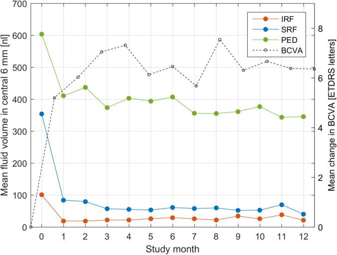
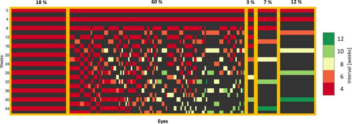
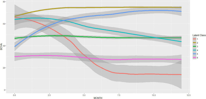
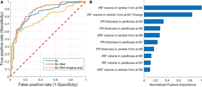
Data of 228 evaluable patients were included, as some had missing scans or were lost to follow-up. Of those patients, 55% reached and maintained long (8, 10, 12 weeks) and another 45% stayed at short (4, 6 weeks) treatment intervals. This provides further evidence for a high disease activity in a major proportion of patients. The model predicted the extendable treatment interval group with an AUROC of 0.71, and the visual outcome with an AUROC of up to 0.87 when utilizing both, clinical and imaging features. The volume of SRF and the volume of IRF, remaining at the first follow-up visit, were found to be the most important predictive markers for treatment intervals and visual outcomes, respectively, supporting the important role of quantitative fluid parameters on OCT.
The proposed Artificial intelligence (AI) methodology was able to predict visual outcomes and retreatment intervals of a T&E regimen from a single injection. The result of this study is an urgently needed step toward AI-supported management of patients with active and progressive nAMD.
使用基于定量光学相干断层扫描(OCT)成像生物标志物的机器学习模型,预测新生血管性年龄相关性黄斑变性(nAMD)的治疗与延长(T&E)方案中的视觉预后和治疗需求。
纳入270例初治受试者的研究眼,这些受试者被随机分配至一项随机临床试验的T&E组接受雷珠单抗治疗。在基线和4周后的首次随访时进行OCT容积扫描。进行自动图像分割,使用卷积神经网络(CNN)勾勒出视网膜内(IRF)、视网膜下(SRF)液、色素上皮脱离(PED)、高反射灶和光感受器层。在早期糖尿病视网膜病变研究(ETDRS)网格上计算一组相应的定量成像生物标志物,以在空间上描述视网膜病理形态及其首次注射后的变化。最后,使用计算出的OCT特征集以及可用的临床和人口统计学信息,使用机器学习建立预后和再治疗间隔的预测模型,并通过10倍交叉验证评估其性能。
纳入了228例可评估患者的数据,因为一些患者扫描缺失或失访。在这些患者中,55%达到并维持了较长(8、10、12周)的治疗间隔,另有45%维持较短(4、6周)的治疗间隔。这为大部分患者的高疾病活动度提供了进一步证据。当同时使用临床和成像特征时,该模型预测可延长治疗间隔组的曲线下面积(AUROC)为0.71,预测视觉预后的AUROC高达0.87。首次随访时残留的SRF体积和IRF体积分别被发现是治疗间隔和视觉预后的最重要预测标志物,支持了OCT上定量液体参数的重要作用。
所提出的人工智能(AI)方法能够从单次注射预测T&E方案的视觉预后和再治疗间隔。本研究结果是朝着AI支持的活动性和进展性nAMD患者管理迈出的迫切需要的一步。