• 文献检索
  • 文档翻译
  • 深度研究
  • 学术资讯
  • Suppr Zotero 插件Zotero 插件
  • 邀请有礼
  • 套餐&价格
  • 历史记录
应用&插件
Suppr Zotero 插件Zotero 插件浏览器插件Mac 客户端Windows 客户端微信小程序
定价
高级版会员购买积分包购买API积分包
服务
文献检索文档翻译深度研究API 文档MCP 服务
关于我们
关于 Suppr公司介绍联系我们用户协议隐私条款
关注我们

Suppr 超能文献

核心技术专利:CN118964589B侵权必究
粤ICP备2023148730 号-1Suppr @ 2026

文献检索

告别复杂PubMed语法,用中文像聊天一样搜索,搜遍4000万医学文献。AI智能推荐,让科研检索更轻松。

立即免费搜索

文件翻译

保留排版,准确专业,支持PDF/Word/PPT等文件格式,支持 12+语言互译。

免费翻译文档

深度研究

AI帮你快速写综述,25分钟生成高质量综述,智能提取关键信息,辅助科研写作。

立即免费体验

MRI 中的联合心室和 WMH 分割,用于评估大脑老化过程中的健康和病理变化。

A joint ventricle and WMH segmentation from MRI for evaluation of healthy and pathological changes in the aging brain.

机构信息

Dept. of Electrical and Computer Engineering, University of Iceland, Reykjavik, Iceland.

Dept. of Medicine, University of Iceland, Reykjavik, Iceland.

出版信息

PLoS One. 2022 Sep 6;17(9):e0274212. doi: 10.1371/journal.pone.0274212. eCollection 2022.

DOI:10.1371/journal.pone.0274212
PMID:36067136
原文链接:https://pmc.ncbi.nlm.nih.gov/articles/PMC9447923/
Abstract

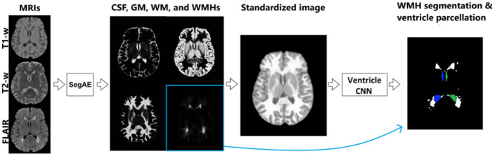
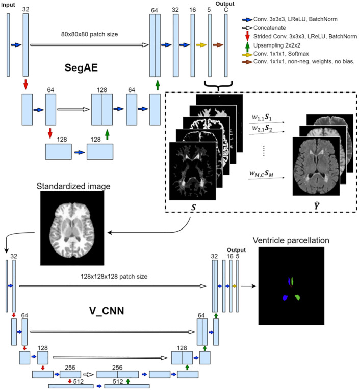
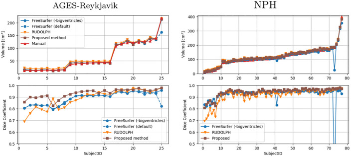
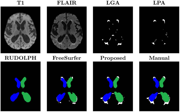
Age-related changes in brain structure include atrophy of the brain parenchyma and white matter changes of presumed vascular origin. Enlargement of the ventricles may occur due to atrophy or impaired cerebrospinal fluid (CSF) circulation. The co-occurrence of these changes in neurodegenerative diseases and in aging brains often requires investigators to take both into account when studying the brain, however, automated segmentation of enlarged ventricles and white matter hyperintensities (WMHs) can be a challenging task. Here, we present a hybrid multi-atlas segmentation and convolutional autoencoder approach for joint ventricle parcellation and WMH segmentation from magnetic resonance images (MRIs). Our fully automated approach uses a convolutional autoencoder to generate a standardized image of grey matter, white matter, CSF, and WMHs, which, in conjunction with labels generated by a multi-atlas segmentation approach, is then fed into a convolutional neural network to parcellate the ventricular system. Hence, our approach does not depend on manually delineated training data for new data sets. The segmentation pipeline was validated on both healthy elderly subjects and subjects with normal pressure hydrocephalus using ground truth manual labels and compared with state-of-the-art segmentation methods. We then applied the method to a cohort of 2401 elderly brains to investigate associations of ventricle volume and WMH load with various demographics and clinical biomarkers, using a multiple regression model. Our results indicate that the ventricle volume and WMH load are both highly variable in a cohort of elderly subjects and there is an independent association between the two, which highlights the importance of taking both the possibility of enlarged ventricles and WMHs into account when studying the aging brain.

摘要

脑结构的年龄相关性变化包括脑实质和可能由血管原因引起的白质变化的萎缩。由于萎缩或脑脊液 (CSF) 循环受损,脑室可能会扩大。在神经退行性疾病和衰老大脑中这些变化的共同发生通常要求研究人员在研究大脑时同时考虑到这两个方面,但是,自动分割扩大的脑室和白质高信号 (WMHs) 可能是一项具有挑战性的任务。在这里,我们提出了一种混合多图谱分割和卷积自动编码器方法,用于从磁共振图像 (MRI) 联合分割脑室和 WMH。我们的全自动方法使用卷积自动编码器生成灰质、白质、CSF 和 WMH 的标准化图像,然后与多图谱分割方法生成的标签结合,输入到卷积神经网络中分割脑室系统。因此,我们的方法不依赖于新数据集的手动描绘的训练数据。该分割管道使用地面真实手动标签在健康老年受试者和正常压力脑积水受试者上进行了验证,并与最先进的分割方法进行了比较。然后,我们使用多元回归模型将该方法应用于 2401 例老年脑队列,以研究脑室体积和 WMH 负荷与各种人口统计学和临床生物标志物之间的关联。我们的结果表明,在老年受试者队列中,脑室体积和 WMH 负荷都具有高度的可变性,并且两者之间存在独立的关联,这突出了在研究衰老大脑时考虑扩大的脑室和 WMH 的可能性的重要性。

https://cdn.ncbi.nlm.nih.gov/pmc/blobs/8e2f/9447923/6079eca67840/pone.0274212.g010.jpg
https://cdn.ncbi.nlm.nih.gov/pmc/blobs/8e2f/9447923/87182a99ef67/pone.0274212.g001.jpg
https://cdn.ncbi.nlm.nih.gov/pmc/blobs/8e2f/9447923/40fc4fb7270c/pone.0274212.g002.jpg
https://cdn.ncbi.nlm.nih.gov/pmc/blobs/8e2f/9447923/9c36580d1b50/pone.0274212.g003.jpg
https://cdn.ncbi.nlm.nih.gov/pmc/blobs/8e2f/9447923/5dbc158c29e3/pone.0274212.g004.jpg
https://cdn.ncbi.nlm.nih.gov/pmc/blobs/8e2f/9447923/858502aa4cd3/pone.0274212.g005.jpg
https://cdn.ncbi.nlm.nih.gov/pmc/blobs/8e2f/9447923/4d84a2e965fc/pone.0274212.g006.jpg
https://cdn.ncbi.nlm.nih.gov/pmc/blobs/8e2f/9447923/e1e04387ff84/pone.0274212.g007.jpg
https://cdn.ncbi.nlm.nih.gov/pmc/blobs/8e2f/9447923/8f2540adac9c/pone.0274212.g008.jpg
https://cdn.ncbi.nlm.nih.gov/pmc/blobs/8e2f/9447923/c5aa677ccf2f/pone.0274212.g009.jpg
https://cdn.ncbi.nlm.nih.gov/pmc/blobs/8e2f/9447923/6079eca67840/pone.0274212.g010.jpg

相似文献

1
A joint ventricle and WMH segmentation from MRI for evaluation of healthy and pathological changes in the aging brain.MRI 中的联合心室和 WMH 分割,用于评估大脑老化过程中的健康和病理变化。
PLoS One. 2022 Sep 6;17(9):e0274212. doi: 10.1371/journal.pone.0274212. eCollection 2022.
2
Evaluation of a deep learning approach for the segmentation of brain tissues and white matter hyperintensities of presumed vascular origin in MRI.基于 MRI 的脑组织结构和可能由血管原因引起的脑白质高信号分割的深度学习方法评估。
Neuroimage Clin. 2017 Oct 12;17:251-262. doi: 10.1016/j.nicl.2017.10.007. eCollection 2018.
3
SegAE: Unsupervised white matter lesion segmentation from brain MRIs using a CNN autoencoder.SegAE:基于 CNN 自动编码器的脑 MRI 无监督白质病变分割。
Neuroimage Clin. 2019;24:102085. doi: 10.1016/j.nicl.2019.102085. Epub 2019 Nov 9.
4
Automatic segmentation of white matter hyperintensities: validation and comparison with state-of-the-art methods on both Multiple Sclerosis and elderly subjects.自动分割脑白质高信号:在多发性硬化症和老年患者中验证和比较最先进的方法。
Neuroimage Clin. 2022;33:102940. doi: 10.1016/j.nicl.2022.102940. Epub 2022 Jan 10.
5
White matter hyperintensity and stroke lesion segmentation and differentiation using convolutional neural networks.基于卷积神经网络的脑白质高信号和卒中病灶分割与区分。
Neuroimage Clin. 2017 Dec 20;17:918-934. doi: 10.1016/j.nicl.2017.12.022. eCollection 2018.
6
BIANCA (Brain Intensity AbNormality Classification Algorithm): A new tool for automated segmentation of white matter hyperintensities.BIANCA(脑强度异常分类算法):一种用于白质高信号自动分割的新工具。
Neuroimage. 2016 Nov 1;141:191-205. doi: 10.1016/j.neuroimage.2016.07.018. Epub 2016 Jul 9.
7
White matter hyperintensity load is associated with premature brain aging.脑白质高信号负荷与脑老化加速有关。
Aging (Albany NY). 2022 Nov 30;14(23):9458-9465. doi: 10.18632/aging.204397.
8
Automatic quantification of white matter hyperintensities on T2-weighted fluid attenuated inversion recovery magnetic resonance imaging.基于 T2 加权液体衰减反转恢复磁共振成像的脑白质高信号自动量化。
Magn Reson Imaging. 2022 Jan;85:71-79. doi: 10.1016/j.mri.2021.10.007. Epub 2021 Oct 16.
9
DEWS (DEep White matter hyperintensity Segmentation framework): A fully automated pipeline for detecting small deep white matter hyperintensities in migraineurs.DEWS(深部白质高信号分割框架):一种用于检测偏头痛患者小深部白质高信号的全自动流水线。
Neuroimage Clin. 2018 Mar 2;18:638-647. doi: 10.1016/j.nicl.2018.02.033. eCollection 2018.
10
Contribution of white matter hyperintensities to ventricular enlargement in older adults.脑白质高信号对老年人脑室扩大的影响。
Neuroimage Clin. 2022;34:103019. doi: 10.1016/j.nicl.2022.103019. Epub 2022 Apr 26.

引用本文的文献

1
Region-based U-nets for fast, accurate, and scalable deep brain segmentation: Application to Parkinson Plus Syndromes.用于快速、准确且可扩展的深部脑部分割的基于区域的U型网络:在帕金森叠加综合征中的应用
Neuroimage Clin. 2025 Jun 24;47:103807. doi: 10.1016/j.nicl.2025.103807.
2
Automated fingerprint analysis as a diagnostic tool for the genetic disorder Kabuki syndrome.自动指纹分析作为遗传性疾病歌舞伎综合征的诊断工具。
Genet Med Open. 2024 Aug 7;2:101884. doi: 10.1016/j.gimo.2024.101884. eCollection 2024.
3
CSF levels of brain-derived proteins correlate with brain ventricular volume in cognitively healthy 70-year-olds.

本文引用的文献

1
Generalizing deep learning brain segmentation for skull removal and intracranial measurements.深度学习脑分割的泛化用于颅骨去除和颅内测量。
Magn Reson Imaging. 2022 May;88:44-52. doi: 10.1016/j.mri.2022.01.004. Epub 2022 Jan 7.
2
Improved Segmentation of the Intracranial and Ventricular Volumes in Populations with Cerebrovascular Lesions and Atrophy Using 3D CNNs.使用 3D CNN 提高脑血管病变和萎缩患者颅内和脑室容积的分割。
Neuroinformatics. 2021 Oct;19(4):597-618. doi: 10.1007/s12021-021-09510-1. Epub 2021 Feb 1.
3
Simultaneous lesion and brain segmentation in multiple sclerosis using deep neural networks.
在认知健康的70岁老人中,脑脊液中脑源性蛋白水平与脑室容积相关。
Clin Proteomics. 2024 Dec 12;21(1):65. doi: 10.1186/s12014-024-09517-1.
4
Comprehensive Overview of Computational Modeling and Artificial Intelligence in Pediatric Neurosurgery.小儿神经外科学中的计算建模与人工智能综合概述。
Adv Exp Med Biol. 2024;1462:487-498. doi: 10.1007/978-3-031-64892-2_30.
5
Specialized gray matter segmentation via a generative adversarial network: application on brain white matter hyperintensities classification.通过生成对抗网络进行的专门灰质分割:在脑白质高信号分类中的应用。
Front Neurosci. 2024 Sep 30;18:1416174. doi: 10.3389/fnins.2024.1416174. eCollection 2024.
6
Investigation of probability maps in deep-learning-based brain ventricle parcellation.基于深度学习的脑室分割中概率图的研究。
Proc SPIE Int Soc Opt Eng. 2023 Feb;12464. doi: 10.1117/12.2653999. Epub 2023 Apr 3.
利用深度神经网络对多发性硬化症进行同时病变和脑区分割。
Sci Rep. 2021 Jan 13;11(1):1087. doi: 10.1038/s41598-020-79925-4.
4
Multi-Domain Image Completion for Random Missing Input Data.多领域图像补全随机缺失输入数据。
IEEE Trans Med Imaging. 2021 Apr;40(4):1113-1122. doi: 10.1109/TMI.2020.3046444. Epub 2021 Apr 1.
5
Shortcomings of Ventricle Segmentation Using Deep Convolutional Networks.使用深度卷积网络进行心室分割的缺点
Underst Interpret Mach Learn Med Image Comput Appl (2018). 2018 Sep;11038:79-86. doi: 10.1007/978-3-030-02628-8_9. Epub 2018 Oct 24.
6
Brain ventricular enlargement in human and murine acute intermittent porphyria.人脑和鼠脑的急性间歇性卟啉症脑室扩大。
Hum Mol Genet. 2020 Nov 25;29(19):3211-3223. doi: 10.1093/hmg/ddaa204.
7
Evaluating White Matter Lesion Segmentations with Refined Sørensen-Dice Analysis.使用改进的 Sørensen-Dice 分析评估脑白质病变分割。
Sci Rep. 2020 May 19;10(1):8242. doi: 10.1038/s41598-020-64803-w.
8
Embracing imperfect datasets: A review of deep learning solutions for medical image segmentation.拥抱不完美数据集:医学图像分割深度学习解决方案综述。
Med Image Anal. 2020 Jul;63:101693. doi: 10.1016/j.media.2020.101693. Epub 2020 Apr 3.
9
SegAE: Unsupervised white matter lesion segmentation from brain MRIs using a CNN autoencoder.SegAE:基于 CNN 自动编码器的脑 MRI 无监督白质病变分割。
Neuroimage Clin. 2019;24:102085. doi: 10.1016/j.nicl.2019.102085. Epub 2019 Nov 9.
10
Ageing as a risk factor for neurodegenerative disease.衰老作为神经退行性疾病的一个风险因素。
Nat Rev Neurol. 2019 Oct;15(10):565-581. doi: 10.1038/s41582-019-0244-7. Epub 2019 Sep 9.