• 文献检索
  • 文档翻译
  • 深度研究
  • 学术资讯
  • Suppr Zotero 插件Zotero 插件
  • 邀请有礼
  • 套餐&价格
  • 历史记录
应用&插件
Suppr Zotero 插件Zotero 插件浏览器插件Mac 客户端Windows 客户端微信小程序
定价
高级版会员购买积分包购买API积分包
服务
文献检索文档翻译深度研究API 文档MCP 服务
关于我们
关于 Suppr公司介绍联系我们用户协议隐私条款
关注我们

Suppr 超能文献

核心技术专利:CN118964589B侵权必究
粤ICP备2023148730 号-1Suppr @ 2026

文献检索

告别复杂PubMed语法,用中文像聊天一样搜索,搜遍4000万医学文献。AI智能推荐,让科研检索更轻松。

立即免费搜索

文件翻译

保留排版,准确专业,支持PDF/Word/PPT等文件格式,支持 12+语言互译。

免费翻译文档

深度研究

AI帮你快速写综述,25分钟生成高质量综述,智能提取关键信息,辅助科研写作。

立即免费体验

利用深度神经网络对多发性硬化症进行同时病变和脑区分割。

Simultaneous lesion and brain segmentation in multiple sclerosis using deep neural networks.

机构信息

Support Center for Advanced Neuroimaging, University Institute for Diagnostic and Interventional Neuroradiology, Inselspital, Bern University Hospital, Bern, Switzerland.

Swiss Paraplegic Centre, Nottwil, Switzerland.

出版信息

Sci Rep. 2021 Jan 13;11(1):1087. doi: 10.1038/s41598-020-79925-4.

DOI:10.1038/s41598-020-79925-4
PMID:33441684
原文链接:https://pmc.ncbi.nlm.nih.gov/articles/PMC7806997/
Abstract

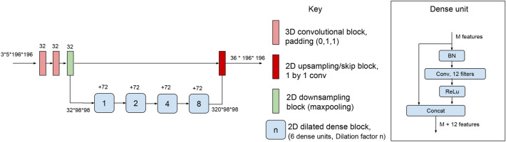
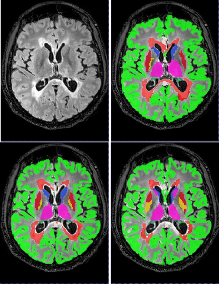
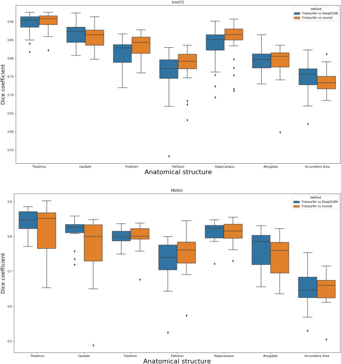
Segmentation of white matter lesions and deep grey matter structures is an important task in the quantification of magnetic resonance imaging in multiple sclerosis. In this paper we explore segmentation solutions based on convolutional neural networks (CNNs) for providing fast, reliable segmentations of lesions and grey-matter structures in multi-modal MR imaging, and the performance of these methods when applied to out-of-centre data. We trained two state-of-the-art fully convolutional CNN architectures on the 2016 MSSEG training dataset, which was annotated by seven independent human raters: a reference implementation of a 3D Unet, and a more recently proposed 3D-to-2D architecture (DeepSCAN). We then retrained those methods on a larger dataset from a single centre, with and without labels for other brain structures. We quantified changes in performance owing to dataset shift, and changes in performance by adding the additional brain-structure labels. We also compared performance with freely available reference methods. Both fully-convolutional CNN methods substantially outperform other approaches in the literature when trained and evaluated in cross-validation on the MSSEG dataset, showing agreement with human raters in the range of human inter-rater variability. Both architectures showed drops in performance when trained on single-centre data and tested on the MSSEG dataset. When trained with the addition of weak anatomical labels derived from Freesurfer, the performance of the 3D Unet degraded, while the performance of the DeepSCAN net improved. Overall, the DeepSCAN network predicting both lesion and anatomical labels was the best-performing network examined.

摘要

对磁共振成像中的白质病变和深部灰质结构进行分割是多发性硬化症量化分析的一项重要任务。在本文中,我们探索了基于卷积神经网络(CNN)的分割解决方案,以便为多模态磁共振成像中的病变和灰质结构提供快速、可靠的分割,并研究这些方法应用于中心外数据时的性能。我们在 2016 年 MSSEG 训练数据集上对两个最先进的全卷积 CNN 架构进行了训练,该数据集由七位独立的人类评估者进行了注释:3D Unet 的参考实现和最近提出的 3D-to-2D 架构(DeepSCAN)。然后,我们在一个来自单一中心的更大数据集上重新训练了这些方法,其中包括或不包括其他脑结构的标签。我们量化了由于数据集偏移和添加其他脑结构标签而导致的性能变化。我们还将性能与免费提供的参考方法进行了比较。在 MSSEG 数据集上进行交叉验证时,这两种全卷积 CNN 方法在训练和评估时都明显优于文献中的其他方法,与人类评估者的一致性在人类评估者之间的可变性范围内。当在单中心数据上进行训练并在 MSSEG 数据集上进行测试时,两种架构的性能都有所下降。当使用来自 Freesurfer 的弱解剖标签进行训练时,3D Unet 的性能下降,而 DeepSCAN 网络的性能提高。总体而言,预测病变和解剖标签的 DeepSCAN 网络是表现最佳的网络。

https://cdn.ncbi.nlm.nih.gov/pmc/blobs/8f2c/7806997/29bbbf3ec168/41598_2020_79925_Fig4_HTML.jpg
https://cdn.ncbi.nlm.nih.gov/pmc/blobs/8f2c/7806997/47c8ae6c01b2/41598_2020_79925_Fig1_HTML.jpg
https://cdn.ncbi.nlm.nih.gov/pmc/blobs/8f2c/7806997/e15839cf01fd/41598_2020_79925_Fig2_HTML.jpg
https://cdn.ncbi.nlm.nih.gov/pmc/blobs/8f2c/7806997/6118eede6ad7/41598_2020_79925_Fig3_HTML.jpg
https://cdn.ncbi.nlm.nih.gov/pmc/blobs/8f2c/7806997/29bbbf3ec168/41598_2020_79925_Fig4_HTML.jpg

相似文献

1
Simultaneous lesion and brain segmentation in multiple sclerosis using deep neural networks.利用深度神经网络对多发性硬化症进行同时病变和脑区分割。
Sci Rep. 2021 Jan 13;11(1):1087. doi: 10.1038/s41598-020-79925-4.
2
Infratentorial lesions in multiple sclerosis patients: intra- and inter-rater variability in comparison to a fully automated segmentation using 3D convolutional neural networks.多发性硬化症患者的桥脑下病变:与使用 3D 卷积神经网络的全自动分割相比的组内和组间可变性。
Eur Radiol. 2022 Apr;32(4):2798-2809. doi: 10.1007/s00330-021-08329-3. Epub 2021 Oct 13.
3
Fully automated longitudinal segmentation of new or enlarged multiple sclerosis lesions using 3D convolutional neural networks.使用 3D 卷积神经网络全自动纵向分割新的或扩大的多发性硬化病变。
Neuroimage Clin. 2020;28:102445. doi: 10.1016/j.nicl.2020.102445. Epub 2020 Sep 24.
4
Multi-branch convolutional neural network for multiple sclerosis lesion segmentation.多分支卷积神经网络在多发性硬化病变分割中的应用。
Neuroimage. 2019 Aug 1;196:1-15. doi: 10.1016/j.neuroimage.2019.03.068. Epub 2019 Apr 3.
5
One-shot domain adaptation in multiple sclerosis lesion segmentation using convolutional neural networks.基于卷积神经网络的多发性硬化病变分割中单样本域自适应
Neuroimage Clin. 2019;21:101638. doi: 10.1016/j.nicl.2018.101638. Epub 2018 Dec 10.
6
Investigation of an efficient multi-modal convolutional neural network for multiple sclerosis lesion detection.一种高效多模态卷积神经网络在多发性硬化病变检测中的研究。
Sci Rep. 2023 Nov 30;13(1):21154. doi: 10.1038/s41598-023-48578-4.
7
Improving automated multiple sclerosis lesion segmentation with a cascaded 3D convolutional neural network approach.采用级联3D卷积神经网络方法改进自动多发性硬化病变分割
Neuroimage. 2017 Jul 15;155:159-168. doi: 10.1016/j.neuroimage.2017.04.034. Epub 2017 Apr 19.
8
Automatic segmentation of the spinal cord and intramedullary multiple sclerosis lesions with convolutional neural networks.基于卷积神经网络的脊髓和髓内多发性硬化病变自动分割。
Neuroimage. 2019 Jan 1;184:901-915. doi: 10.1016/j.neuroimage.2018.09.081. Epub 2018 Oct 6.
9
Neuro-fuzzy patch-wise R-CNN for multiple sclerosis segmentation.基于神经模糊补丁 R-CNN 的多发性硬化分割。
Med Biol Eng Comput. 2020 Sep;58(9):2161-2175. doi: 10.1007/s11517-020-02225-6. Epub 2020 Jul 17.
10
A Novel Public MR Image Dataset of Multiple Sclerosis Patients With Lesion Segmentations Based on Multi-rater Consensus.一种基于多位评估者共识的多发性硬化症患者病变分割的新型公共磁共振图像数据集。
Neuroinformatics. 2018 Jan;16(1):51-63. doi: 10.1007/s12021-017-9348-7.

引用本文的文献

1
Revealing morphological fingerprints in perinatal brains using quasi-conformal mapping: occurrence and neurodevelopmental implications.使用拟共形映射揭示围产期大脑中的形态学特征:发生情况及对神经发育的影响
Brain Imaging Behav. 2025 Mar 27. doi: 10.1007/s11682-025-00998-8.
2
Machine learning for refining interpretation of magnetic resonance imaging scans in the management of multiple sclerosis: a narrative review.机器学习在多发性硬化症管理中优化磁共振成像扫描解读的应用:一项叙述性综述
R Soc Open Sci. 2025 Jan 22;12(1):241052. doi: 10.1098/rsos.241052. eCollection 2025 Jan.
3
Diagnostic effectiveness of deep learning-based MRI in predicting multiple sclerosis: A meta-analysis.

本文引用的文献

1
A contrast-adaptive method for simultaneous whole-brain and lesion segmentation in multiple sclerosis.一种用于多发性硬化症的全脑和病灶同时分割的对比自适应方法。
Neuroimage. 2021 Jan 15;225:117471. doi: 10.1016/j.neuroimage.2020.117471. Epub 2020 Oct 22.
2
Multiple sclerosis cortical and WM lesion segmentation at 3T MRI: a deep learning method based on FLAIR and MP2RAGE.基于 FLAIR 和 MP2RAGE 的深度学习方法在 3T MRI 下对多发性硬化症皮质和 WM 病变的分割。
Neuroimage Clin. 2020;27:102335. doi: 10.1016/j.nicl.2020.102335. Epub 2020 Jun 30.
3
Automatic detection of lesion load change in Multiple Sclerosis using convolutional neural networks with segmentation confidence.
基于深度学习的 MRI 在预测多发性硬化症中的诊断效能:一项荟萃分析。
Neurosciences (Riyadh). 2024 May;29(2):77-89. doi: 10.17712/nsj.2024.2.20230103.
4
The impact of image contrast, resolution and reader expertise on black hole identification in Multiple Sclerosis.图像对比度、分辨率和阅片者专业知识对多发性硬化症中黑洞识别的影响。
Neuroradiology. 2024 Aug;66(8):1345-1352. doi: 10.1007/s00234-024-03310-5. Epub 2024 Feb 19.
5
LST-AI: a Deep Learning Ensemble for Accurate MS Lesion Segmentation.LST-AI:用于准确的多发性硬化症病变分割的深度学习集成方法
medRxiv. 2024 Mar 11:2023.11.23.23298966. doi: 10.1101/2023.11.23.23298966.
6
3D U-Net for automated detection of multiple sclerosis lesions: utility of transfer learning from other pathologies.用于自动检测多发性硬化症病变的3D U-Net:从其他病理学进行迁移学习的效用。
Front Neurosci. 2023 Oct 27;17:1188336. doi: 10.3389/fnins.2023.1188336. eCollection 2023.
7
BIANCA-MS: An optimized tool for automated multiple sclerosis lesion segmentation.Bianca-MS:用于自动多发性硬化病变分割的优化工具。
Hum Brain Mapp. 2023 Oct 1;44(14):4893-4913. doi: 10.1002/hbm.26424. Epub 2023 Aug 2.
8
Scanner agnostic large-scale evaluation of MS lesion delineation tool for clinical MRI.临床MRI中MS病变勾画工具的扫描仪无关大规模评估
Front Neurosci. 2023 May 19;17:1177540. doi: 10.3389/fnins.2023.1177540. eCollection 2023.
9
Multi-arm U-Net with dense input and skip connectivity for T2 lesion segmentation in clinical trials of multiple sclerosis.多臂 U-Net 具有密集输入和跳过连接,用于多发性硬化症临床试验中的 T2 病变分割。
Sci Rep. 2023 Mar 13;13(1):4102. doi: 10.1038/s41598-023-31207-5.
10
A joint ventricle and WMH segmentation from MRI for evaluation of healthy and pathological changes in the aging brain.MRI 中的联合心室和 WMH 分割,用于评估大脑老化过程中的健康和病理变化。
PLoS One. 2022 Sep 6;17(9):e0274212. doi: 10.1371/journal.pone.0274212. eCollection 2022.
利用具有分割置信度的卷积神经网络自动检测多发性硬化症的病变负荷变化。
Neuroimage Clin. 2020;25:102104. doi: 10.1016/j.nicl.2019.102104. Epub 2019 Dec 9.
4
Asymmetric Loss Functions and Deep Densely Connected Networks for Highly Imbalanced Medical Image Segmentation: Application to Multiple Sclerosis Lesion Detection.用于高度不平衡医学图像分割的非对称损失函数和深度密集连接网络:在多发性硬化症病变检测中的应用
IEEE Access. 2019;7:721-1735. doi: 10.1109/ACCESS.2018.2886371. Epub 2018 Dec 12.
5
Automated Detection and Segmentation of Multiple Sclerosis Lesions Using Ultra-High-Field MP2RAGE.基于超高场 MP2RAGE 的多发性硬化病变的自动检测和分割。
Invest Radiol. 2019 Jun;54(6):356-364. doi: 10.1097/RLI.0000000000000551.
6
Geostatistical Analysis of White Matter Lesions in Multiple Sclerosis Identifies Gender Differences in Lesion Evolution.多发性硬化症白质病变的地质统计学分析揭示了病变演变中的性别差异。
Front Mol Neurosci. 2018 Dec 17;11:460. doi: 10.3389/fnmol.2018.00460. eCollection 2018.
7
One-shot domain adaptation in multiple sclerosis lesion segmentation using convolutional neural networks.基于卷积神经网络的多发性硬化病变分割中单样本域自适应
Neuroimage Clin. 2019;21:101638. doi: 10.1016/j.nicl.2018.101638. Epub 2018 Dec 10.
8
Predicting conversion from clinically isolated syndrome to multiple sclerosis-An imaging-based machine learning approach.基于影像的机器学习方法预测临床孤立综合征向多发性硬化症的转化。
Neuroimage Clin. 2019;21:101593. doi: 10.1016/j.nicl.2018.11.003. Epub 2018 Nov 5.
9
Repeatability and reproducibility of FreeSurfer, FSL-SIENAX and SPM brain volumetric measurements and the effect of lesion filling in multiple sclerosis.FreeSurfer、FSL-SIENAX 和 SPM 脑容量测量的可重复性和再现性,以及多发性硬化症中病变填充的影响。
Eur Radiol. 2019 Mar;29(3):1355-1364. doi: 10.1007/s00330-018-5710-x. Epub 2018 Sep 21.
10
Objective Evaluation of Multiple Sclerosis Lesion Segmentation using a Data Management and Processing Infrastructure.利用数据管理和处理基础设施对多发性硬化病变进行客观评估。
Sci Rep. 2018 Sep 12;8(1):13650. doi: 10.1038/s41598-018-31911-7.