• 文献检索
  • 文档翻译
  • 深度研究
  • 学术资讯
  • Suppr Zotero 插件Zotero 插件
  • 邀请有礼
  • 套餐&价格
  • 历史记录
应用&插件
Suppr Zotero 插件Zotero 插件浏览器插件Mac 客户端Windows 客户端微信小程序
定价
高级版会员购买积分包购买API积分包
服务
文献检索文档翻译深度研究API 文档MCP 服务
关于我们
关于 Suppr公司介绍联系我们用户协议隐私条款
关注我们

Suppr 超能文献

核心技术专利:CN118964589B侵权必究
粤ICP备2023148730 号-1Suppr @ 2026

文献检索

告别复杂PubMed语法,用中文像聊天一样搜索,搜遍4000万医学文献。AI智能推荐,让科研检索更轻松。

立即免费搜索

文件翻译

保留排版,准确专业,支持PDF/Word/PPT等文件格式,支持 12+语言互译。

免费翻译文档

深度研究

AI帮你快速写综述,25分钟生成高质量综述,智能提取关键信息,辅助科研写作。

立即免费体验

基于 MRI 的脑组织结构和可能由血管原因引起的脑白质高信号分割的深度学习方法评估。

Evaluation of a deep learning approach for the segmentation of brain tissues and white matter hyperintensities of presumed vascular origin in MRI.

机构信息

Image Sciences Institute, University Medical Center Utrecht and Utrecht University, The Netherlands; Medical Image Analysis, Department of Biomedical Engineering, Eindhoven University of Technology, The Netherlands.

Department of Radiology, University Medical Center Utrecht, The Netherlands.

出版信息

Neuroimage Clin. 2017 Oct 12;17:251-262. doi: 10.1016/j.nicl.2017.10.007. eCollection 2018.

DOI:10.1016/j.nicl.2017.10.007
PMID:29159042
原文链接:https://pmc.ncbi.nlm.nih.gov/articles/PMC5683197/
Abstract

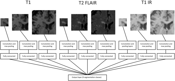
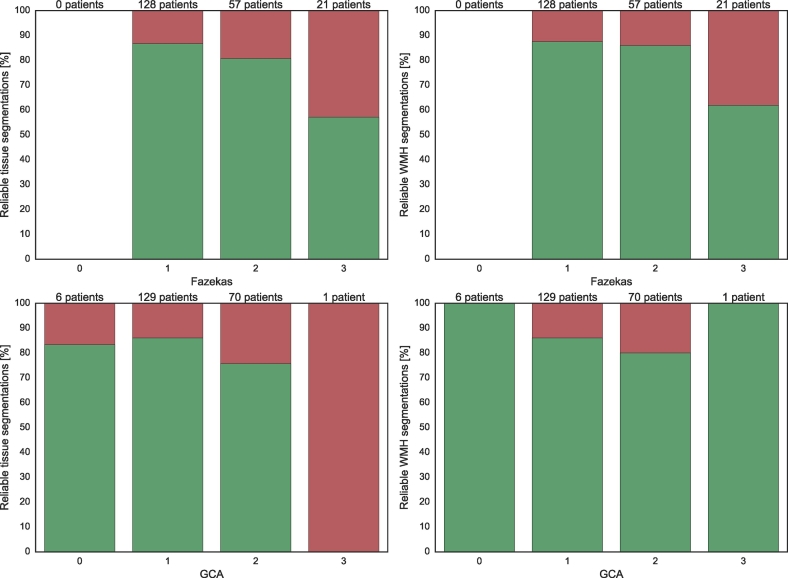
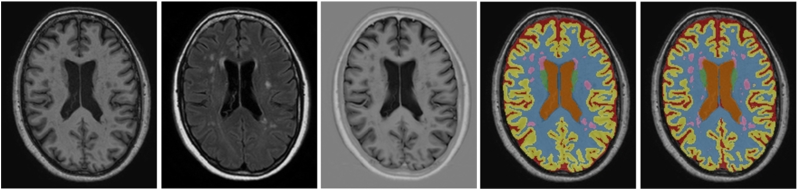
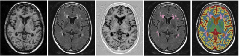
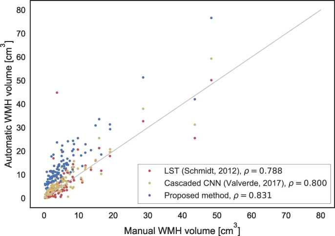
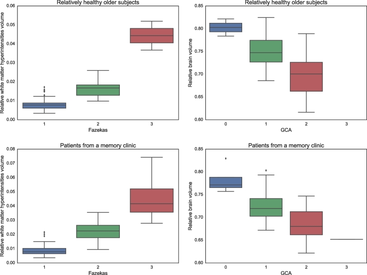
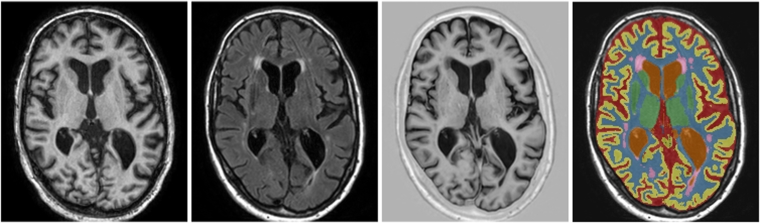
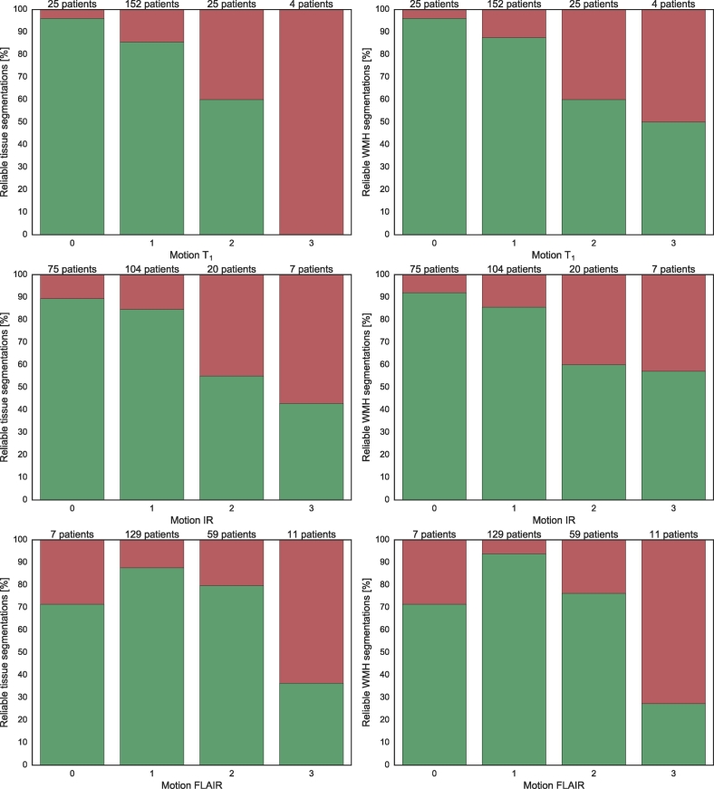
Automatic segmentation of brain tissues and white matter hyperintensities of presumed vascular origin (WMH) in MRI of older patients is widely described in the literature. Although brain abnormalities and motion artefacts are common in this age group, most segmentation methods are not evaluated in a setting that includes these items. In the present study, our tissue segmentation method for brain MRI was extended and evaluated for additional WMH segmentation. Furthermore, our method was evaluated in two large cohorts with a realistic variation in brain abnormalities and motion artefacts. The method uses a multi-scale convolutional neural network with a T-weighted image, a T-weighted fluid attenuated inversion recovery (FLAIR) image and a T-weighted inversion recovery (IR) image as input. The method automatically segments white matter (WM), cortical grey matter (cGM), basal ganglia and thalami (BGT), cerebellum (CB), brain stem (BS), lateral ventricular cerebrospinal fluid (lvCSF), peripheral cerebrospinal fluid (pCSF), and WMH. Our method was evaluated quantitatively with images publicly available from the MRBrainS13 challenge ( = 20), quantitatively and qualitatively in relatively healthy older subjects ( = 96), and qualitatively in patients from a memory clinic ( = 110). The method can accurately segment WMH (Overall Dice coefficient in the MRBrainS13 data of 0.67) without compromising performance for tissue segmentations (Overall Dice coefficients in the MRBrainS13 data of 0.87 for WM, 0.85 for cGM, 0.82 for BGT, 0.93 for CB, 0.92 for BS, 0.93 for lvCSF, 0.76 for pCSF). Furthermore, the automatic WMH volumes showed a high correlation with manual WMH volumes (Spearman's  = 0.83 for relatively healthy older subjects). In both cohorts, our method produced reliable segmentations (as determined by a human observer) in most images (relatively healthy/memory clinic: tissues 88%/77% reliable, WMH 85%/84% reliable) despite various degrees of brain abnormalities and motion artefacts. In conclusion, this study shows that a convolutional neural network-based segmentation method can accurately segment brain tissues and WMH in MR images of older patients with varying degrees of brain abnormalities and motion artefacts.

摘要

在老年患者的 MRI 中,自动分割脑组织结构和由假定血管来源引起的脑白质高信号(WMH)在文献中被广泛描述。尽管在这个年龄段,脑异常和运动伪影很常见,但大多数分割方法并没有在包含这些项目的环境中进行评估。在本研究中,我们扩展了用于脑 MRI 的组织分割方法,并对额外的 WMH 分割进行了评估。此外,我们的方法在具有不同程度脑异常和运动伪影的两个大型队列中进行了评估。该方法使用多尺度卷积神经网络,以 T 加权图像、T 加权液体衰减反转恢复(FLAIR)图像和 T 加权反转恢复(IR)图像作为输入。该方法自动分割脑白质(WM)、皮质灰质(cGM)、基底节和丘脑(BGT)、小脑(CB)、脑桥(BS)、侧脑室脑脊液(lvCSF)、周围脑脊液(pCSF)和 WMH。我们的方法使用公开的 MRBrainS13 挑战赛图像进行了定量评估(=20),使用相对健康的老年受试者进行了定量和定性评估(=96),并使用记忆诊所的患者进行了定性评估(=110)。该方法可以准确分割 WMH(在 MRBrainS13 数据中的总体 Dice 系数为 0.67),而不会影响组织分割的性能(在 MRBrainS13 数据中的总体 Dice 系数为 WM 0.87、cGM 0.85、BGT 0.82、CB 0.93、BS 0.92、lvCSF 0.93、pCSF 0.76)。此外,自动 WMH 体积与手动 WMH 体积具有高度相关性(相对健康的老年受试者的 Spearman's 系数为 0.83)。在两个队列中,尽管存在不同程度的脑异常和运动伪影,但我们的方法在大多数图像中都产生了可靠的分割(由人类观察者确定)(相对健康/记忆诊所:组织 88%/77%可靠,WMH 85%/84%可靠)。总之,本研究表明,基于卷积神经网络的分割方法可以准确分割具有不同程度脑异常和运动伪影的老年患者的脑组织结构和 WMH。

https://cdn.ncbi.nlm.nih.gov/pmc/blobs/0e35/5683197/3dd7f04182b1/gr12.jpg
https://cdn.ncbi.nlm.nih.gov/pmc/blobs/0e35/5683197/ce298232061d/gr1.jpg
https://cdn.ncbi.nlm.nih.gov/pmc/blobs/0e35/5683197/84c50e84ff4e/gr2.jpg
https://cdn.ncbi.nlm.nih.gov/pmc/blobs/0e35/5683197/c4682a3cbb2f/gr3.jpg
https://cdn.ncbi.nlm.nih.gov/pmc/blobs/0e35/5683197/c9436cc48f6d/gr4.jpg
https://cdn.ncbi.nlm.nih.gov/pmc/blobs/0e35/5683197/ee79731941f1/gr5.jpg
https://cdn.ncbi.nlm.nih.gov/pmc/blobs/0e35/5683197/9359c50684ea/gr6.jpg
https://cdn.ncbi.nlm.nih.gov/pmc/blobs/0e35/5683197/f321c4e15573/gr7.jpg
https://cdn.ncbi.nlm.nih.gov/pmc/blobs/0e35/5683197/2e515a11f0a1/gr8.jpg
https://cdn.ncbi.nlm.nih.gov/pmc/blobs/0e35/5683197/7113bad876b4/gr9.jpg
https://cdn.ncbi.nlm.nih.gov/pmc/blobs/0e35/5683197/879fcb5a4da6/gr10.jpg
https://cdn.ncbi.nlm.nih.gov/pmc/blobs/0e35/5683197/5082be12f02a/gr11.jpg
https://cdn.ncbi.nlm.nih.gov/pmc/blobs/0e35/5683197/3dd7f04182b1/gr12.jpg

相似文献

1
Evaluation of a deep learning approach for the segmentation of brain tissues and white matter hyperintensities of presumed vascular origin in MRI.基于 MRI 的脑组织结构和可能由血管原因引起的脑白质高信号分割的深度学习方法评估。
Neuroimage Clin. 2017 Oct 12;17:251-262. doi: 10.1016/j.nicl.2017.10.007. eCollection 2018.
2
Automatic segmentation of white matter hyperintensities: validation and comparison with state-of-the-art methods on both Multiple Sclerosis and elderly subjects.自动分割脑白质高信号:在多发性硬化症和老年患者中验证和比较最先进的方法。
Neuroimage Clin. 2022;33:102940. doi: 10.1016/j.nicl.2022.102940. Epub 2022 Jan 10.
3
UBO Detector - A cluster-based, fully automated pipeline for extracting white matter hyperintensities.UBO 探测器——一种基于聚类的、全自动的提取脑白质高信号的流水线。
Neuroimage. 2018 Jul 1;174:539-549. doi: 10.1016/j.neuroimage.2018.03.050. Epub 2018 Mar 22.
4
Segmentation of white matter hyperintensities using convolutional neural networks with global spatial information in routine clinical brain MRI with none or mild vascular pathology.利用卷积神经网络和全局空间信息对常规临床脑部 MRI(无或轻度血管病变)中的脑白质高信号进行分割。
Comput Med Imaging Graph. 2018 Jun;66:28-43. doi: 10.1016/j.compmedimag.2018.02.002. Epub 2018 Feb 17.
5
Automatic quantification of white matter hyperintensities on T2-weighted fluid attenuated inversion recovery magnetic resonance imaging.基于 T2 加权液体衰减反转恢复磁共振成像的脑白质高信号自动量化。
Magn Reson Imaging. 2022 Jan;85:71-79. doi: 10.1016/j.mri.2021.10.007. Epub 2021 Oct 16.
6
White matter hyperintensity and stroke lesion segmentation and differentiation using convolutional neural networks.基于卷积神经网络的脑白质高信号和卒中病灶分割与区分。
Neuroimage Clin. 2017 Dec 20;17:918-934. doi: 10.1016/j.nicl.2017.12.022. eCollection 2018.
7
Limited One-time Sampling Irregularity Map (LOTS-IM) for Automatic Unsupervised Assessment of White Matter Hyperintensities and Multiple Sclerosis Lesions in Structural Brain Magnetic Resonance Images.用于结构磁共振成像中白质高信号和多发性硬化病变自动无监督评估的有限一次性采样不规则图(LOTS-IM)。
Comput Med Imaging Graph. 2020 Jan;79:101685. doi: 10.1016/j.compmedimag.2019.101685. Epub 2019 Nov 27.
8
A joint ventricle and WMH segmentation from MRI for evaluation of healthy and pathological changes in the aging brain.MRI 中的联合心室和 WMH 分割,用于评估大脑老化过程中的健康和病理变化。
PLoS One. 2022 Sep 6;17(9):e0274212. doi: 10.1371/journal.pone.0274212. eCollection 2022.
9
Improved Automatic Segmentation of White Matter Hyperintensities in MRI Based on Multilevel Lesion Features.基于多级病变特征的磁共振成像中白质高信号的改进自动分割
Neuroinformatics. 2017 Jul;15(3):231-245. doi: 10.1007/s12021-017-9328-y.
10
Metric to quantify white matter damage on brain magnetic resonance images.用于量化脑磁共振图像上白质损伤的指标。
Neuroradiology. 2017 Oct;59(10):951-962. doi: 10.1007/s00234-017-1892-1. Epub 2017 Aug 16.

引用本文的文献

1
Enhanced Brain Stroke Lesion Segmentation in MRI Using a 2.5D Transformer Backbone U-Net Model.使用2.5D变压器主干U-Net模型增强MRI中的脑卒中标注分割
Brain Sci. 2025 Jul 22;15(8):778. doi: 10.3390/brainsci15080778.
2
Segmentation of white matter lesions in multicentre FLAIR MRI.多中心液体衰减反转恢复磁共振成像(FLAIR MRI)中白质病变的分割
Neuroimage Rep. 2021 Aug 5;1(4):100044. doi: 10.1016/j.ynirp.2021.100044. eCollection 2021 Dec.
3
Artificial intelligence for volumetric measurement of cerebral white matter hyperintensities on thick-slice fluid-attenuated inversion recovery (FLAIR) magnetic resonance images from multiple centers.

本文引用的文献

1
Location Sensitive Deep Convolutional Neural Networks for Segmentation of White Matter Hyperintensities.位置敏感深度卷积神经网络在脑白质高信号分割中的应用
Sci Rep. 2017 Jul 11;7(1):5110. doi: 10.1038/s41598-017-05300-5.
2
Improving automated multiple sclerosis lesion segmentation with a cascaded 3D convolutional neural network approach.采用级联3D卷积神经网络方法改进自动多发性硬化病变分割
Neuroimage. 2017 Jul 15;155:159-168. doi: 10.1016/j.neuroimage.2017.04.034. Epub 2017 Apr 19.
3
Deep multi-scale location-aware 3D convolutional neural networks for automated detection of lacunes of presumed vascular origin.
多中心厚层液体衰减反转恢复(FLAIR)磁共振图像脑白质高信号容积测量的人工智能。
Sci Rep. 2024 May 2;14(1):10104. doi: 10.1038/s41598-024-60789-x.
4
Utilizing deep learning via the 3D U-net neural network for the delineation of brain stroke lesions in MRI image.利用 3D U-net 神经网络的深度学习对 MRI 图像中的脑卒中风病变进行勾画。
Sci Rep. 2023 Nov 13;13(1):19808. doi: 10.1038/s41598-023-47107-7.
5
Artificial Intelligence in Neuroradiology: A Review of Current Topics and Competition Challenges.神经放射学中的人工智能:当前主题与竞争挑战综述
Diagnostics (Basel). 2023 Aug 14;13(16):2670. doi: 10.3390/diagnostics13162670.
6
Toward attention-based learning to predict the risk of brain degeneration with multimodal medical data.迈向基于注意力的学习,以利用多模态医学数据预测脑退化风险。
Front Neurosci. 2023 Jan 18;16:1043626. doi: 10.3389/fnins.2022.1043626. eCollection 2022.
7
Semisupervised white matter hyperintensities segmentation on MRI.基于半监督学习的 MRI 脑白质高信号分割。
Hum Brain Mapp. 2023 Mar;44(4):1344-1358. doi: 10.1002/hbm.26109. Epub 2022 Oct 10.
8
Ensemble learning supervision augmentation for white matter hyperintensity segmentation.用于白质高信号分割的集成学习监督增强
Front Neurosci. 2022 Sep 15;16:946343. doi: 10.3389/fnins.2022.946343. eCollection 2022.
9
Diagnostic performance of deep learning-based automatic white matter hyperintensity segmentation for classification of the Fazekas scale and differentiation of subcortical vascular dementia.基于深度学习的自动脑白质高信号分割对 Fazekas 量表分类和皮质下血管性痴呆鉴别诊断的性能评估。
PLoS One. 2022 Sep 15;17(9):e0274562. doi: 10.1371/journal.pone.0274562. eCollection 2022.
10
Automatic segmentation of white matter hyperintensities in routine clinical brain MRI by 2D VB-Net: A large-scale study.基于二维VB-Net的常规临床脑部MRI中白质高信号自动分割:一项大规模研究。
Front Aging Neurosci. 2022 Jul 29;14:915009. doi: 10.3389/fnagi.2022.915009. eCollection 2022.
用于自动检测假定血管源性腔隙的深度多尺度位置感知3D卷积神经网络
Neuroimage Clin. 2017 Feb 4;14:391-399. doi: 10.1016/j.nicl.2017.01.033. eCollection 2017.
4
Automated detection of white matter hyperintensities of all sizes in cerebral small vessel disease.脑小血管病中各种大小白质高信号的自动检测
Med Phys. 2016 Dec;43(12):6246. doi: 10.1118/1.4966029.
5
Efficient multi-scale 3D CNN with fully connected CRF for accurate brain lesion segmentation.高效多尺度 3D CNN 结合全连接条件随机场实现精准脑损伤分割。
Med Image Anal. 2017 Feb;36:61-78. doi: 10.1016/j.media.2016.10.004. Epub 2016 Oct 29.
6
Robustness of Automated Methods for Brain Volume Measurements across Different MRI Field Strengths.不同MRI场强下脑容量测量自动化方法的稳健性
PLoS One. 2016 Oct 31;11(10):e0165719. doi: 10.1371/journal.pone.0165719. eCollection 2016.
7
Brain tumor segmentation with Deep Neural Networks.基于深度神经网络的脑肿瘤分割。
Med Image Anal. 2017 Jan;35:18-31. doi: 10.1016/j.media.2016.05.004. Epub 2016 May 19.
8
Automatic Segmentation of MR Brain Images With a Convolutional Neural Network.基于卷积神经网络的磁共振脑图像自动分割。
IEEE Trans Med Imaging. 2016 May;35(5):1252-1261. doi: 10.1109/TMI.2016.2548501. Epub 2016 Mar 30.
9
Brain Tumor Segmentation Using Convolutional Neural Networks in MRI Images.基于 MRI 图像的卷积神经网络脑肿瘤分割。
IEEE Trans Med Imaging. 2016 May;35(5):1240-1251. doi: 10.1109/TMI.2016.2538465. Epub 2016 Mar 4.
10
Deep 3D Convolutional Encoder Networks With Shortcuts for Multiscale Feature Integration Applied to Multiple Sclerosis Lesion Segmentation.具有捷径的深层 3D 卷积编码网络,用于多尺度特征集成,应用于多发性硬化病变分割。
IEEE Trans Med Imaging. 2016 May;35(5):1229-1239. doi: 10.1109/TMI.2016.2528821. Epub 2016 Feb 11.